2CY3

CRYSTAL STRUCTURE OF CYTOCHROME C3 FROM DESULFOVIBRIO DESULFURICANS NORWAY AT 1.7 ANGSTROMS RESOLUTION


Experimental Data Snapshot

  • Method: X-RAY DIFFRACTION
  • Resolution: 1.70 Å
  • R-Value Work: 0.198 
  • R-Value Observed: 0.198 

wwPDB Validation   3D Report Full Report


This is version 2.0 of the entry. See complete history


Literature

Crystal structure of cytochrome c3 from Desulfovibrio desulfuricans Norway at 1.7 A resolution.

Czjzek, M.Payan, F.Guerlesquin, F.Bruschi, M.Haser, R.

(1994) J Mol Biol 243: 653-667

  • DOI: https://doi.org/10.1016/0022-2836(94)90039-6
  • Primary Citation of Related Structures:  
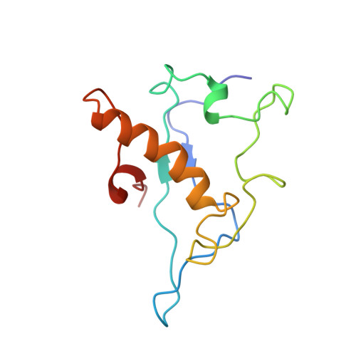
    2CY3

  • PubMed Abstract: 

    The crystal structure of cytochrome c3 (M(r) 13,000) from Desulfovibrio desulfuricans (118 residues, four heme groups) has been crystallographically refined to 1.7 A resolution using a simulated annealing method, based on the structure-model at 2.5 A resolution, already published. The final R-factor for 10,549 reflections was 0.198 covering the range from 5.5 to 1.7 A resolution. The individual temperature factors were refined for a total of 1059 protein atoms, together with 126 bound solvent molecules. The structure has been analyzed with respect to its detailed conformational properties, secondary structure features, temperature factor behaviour, bound solvent sites and heme geometry and ligation. The characteristic secondary structures of the polypeptide chain of this molecule are one extended alpha-helix, a short beta-strand and 13 reverse turns. The four heme groups are located in different structural environments, all highly exposed to solvent. The particular structural features of the heme environments are compared to the four hemes of the cytochrome c3 from Desulfovibrio vulgaris Miyazaki.


  • Organizational Affiliation

    CNRS-Marseille, Laboratoire de Cristallographie et Cristallisation des Macromolécules Biologiques, URA 1296 Faculté de Médicine-Nord, Marseille, France.


Macromolecules
Find similar proteins by:  (by identity cutoff)  |  3D Structure
Entity ID: 1
MoleculeChains Sequence LengthOrganismDetailsImage
CYTOCHROME C3118Desulfomicrobium norvegicumMutation(s): 0 
UniProt
Find proteins for P00136 (Desulfomicrobium norvegicum (strain DSM 1741 / NCIMB 8310))
Explore P00136 
Go to UniProtKB:  P00136
Entity Groups  
Sequence Clusters30% Identity50% Identity70% Identity90% Identity95% Identity100% Identity
UniProt GroupP00136
Sequence Annotations
Expand
  • Reference Sequence
Experimental Data & Validation

Experimental Data

  • Method: X-RAY DIFFRACTION
  • Resolution: 1.70 Å
  • R-Value Work: 0.198 
  • R-Value Observed: 0.198 
  • Space Group: P 31
Unit Cell:
Length ( Å )Angle ( ˚ )
a = 43.72α = 90
b = 43.72β = 90
c = 64.45γ = 120
Software Package:
Software NamePurpose
X-PLORmodel building
X-PLORrefinement
X-PLORphasing

Structure Validation

View Full Validation Report


Entry History 

Deposition Data

Revision History  (Full details and data files)

  • Version 1.0: 1994-10-15
    Type: Initial release
  • Version 1.1: 2008-03-24
    Changes: Version format compliance
  • Version 1.2: 2011-07-13
    Changes: Version format compliance
  • Version 1.3: 2017-11-29
    Changes: Derived calculations, Other
  • Version 2.0: 2021-03-03
    Changes: Advisory, Atomic model, Data collection, Derived calculations, Non-polymer description, Structure summary