2CG6

Second and third fibronectin type I module pair (crystal form I).


Experimental Data Snapshot

  • Method: X-RAY DIFFRACTION
  • Resolution: 1.55 Å
  • R-Value Free: 0.231 
  • R-Value Work: 0.188 
  • R-Value Observed: 0.191 

wwPDB Validation   3D Report Full Report


This is version 1.3 of the entry. See complete history


Literature

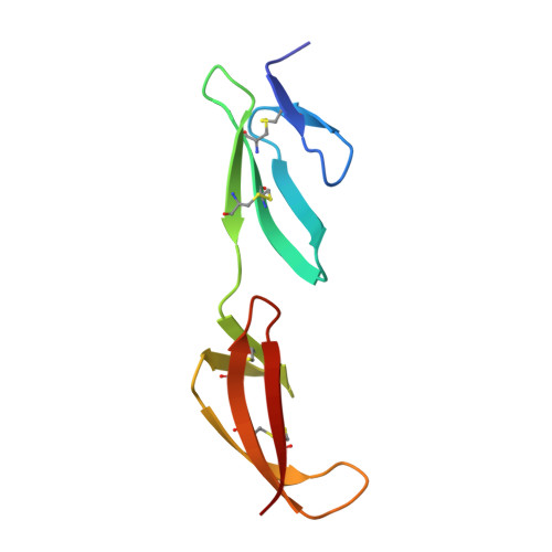
The Solution and Crystal Structures of a Module Pair from the Staphylococcus Aureus-Binding Site of Human Fibronectin-A Tale with a Twist.

Rudino-Pinera, E.Ravelli, R.B.G.Sheldrick, G.M.Nanao, M.H.Korostelev, V.V.Werner, J.M.Schwarz-Linek, U.Potts, J.R.Garman, E.F.

(2007) J Mol Biol 368: 833

  • DOI: https://doi.org/10.1016/j.jmb.2007.02.061
  • Primary Citation of Related Structures:  
    2CG6, 2CG7, 2CKU

  • PubMed Abstract: 

    An important goal of structural studies of modular proteins is to determine the inter-module orientation, which often influences biological function. The N-terminal domain of human fibronectin (Fn) is composed of a string of five type 1 modules (F1). Despite their small size, to date F1 modules have proved intractable to X-ray structure solution, although there are several NMR structures available. Here, we present the first structures (two X-ray models and an NMR-derived model) of the (2)F1(3)F1 module pair, which forms part of the binding site for Fn-binding proteins from pathogenic bacteria. The crystallographic structure determination was aided by the novel technique of UV radiation damage-induced phasing. The individual module structures are very similar in all three models. In the NMR structure and one of the X-ray structures, a similar but smaller interdomain interface than that observed previously for (4)F1(5)F1 is seen. The other X-ray structure has a different interdomain orientation. This work underlines the benefits of combining X-ray and NMR data in the studies of multi-domain proteins.


  • Organizational Affiliation

    Department of Biochemistry, University of Oxford, South Parks Road, Oxford, UK.


Macromolecules
Find similar proteins by:  (by identity cutoff)  |  3D Structure
Entity ID: 1
MoleculeChains Sequence LengthOrganismDetailsImage
HUMAN FIBRONECTIN90Homo sapiensMutation(s): 0 
UniProt & NIH Common Fund Data Resources
Find proteins for P02751 (Homo sapiens)
Explore P02751 
Go to UniProtKB:  P02751
PHAROS:  P02751
GTEx:  ENSG00000115414 
Entity Groups  
Sequence Clusters30% Identity50% Identity70% Identity90% Identity95% Identity100% Identity
UniProt GroupP02751
Sequence Annotations
Expand
  • Reference Sequence
Small Molecules
Ligands 1 Unique
IDChains Name / Formula / InChI Key2D Diagram3D Interactions
SO4
Query on SO4

Download Ideal Coordinates CCD File 
B [auth A]SULFATE ION
O4 S
QAOWNCQODCNURD-UHFFFAOYSA-L
Experimental Data & Validation

Experimental Data

  • Method: X-RAY DIFFRACTION
  • Resolution: 1.55 Å
  • R-Value Free: 0.231 
  • R-Value Work: 0.188 
  • R-Value Observed: 0.191 
  • Space Group: P 41 21 2
Unit Cell:
Length ( Å )Angle ( ˚ )
a = 37.855α = 90
b = 37.855β = 90
c = 108.419γ = 90
Software Package:
Software NamePurpose
REFMACrefinement
MOSFLMdata reduction
SCALAdata scaling
SHELXphasing

Structure Validation

View Full Validation Report


Entry History 

Deposition Data

Revision History  (Full details and data files)

  • Version 1.0: 2007-02-27
    Type: Initial release
  • Version 1.1: 2011-05-08
    Changes: Version format compliance
  • Version 1.2: 2011-07-13
    Changes: Version format compliance
  • Version 1.3: 2017-08-30
    Changes: Data collection