Refined structure of cytochrome c3 at 1.8 A resolution
Higuchi, Y., Kusunoki, M., Matsuura, Y., Yasuoka, N., Kakudo, M.(1984) J Mol Biol 172: 109-139
- PubMed: 6319712
- DOI: https://doi.org/10.1016/0022-2836(84)90417-0
- Primary Citation of Related Structures:
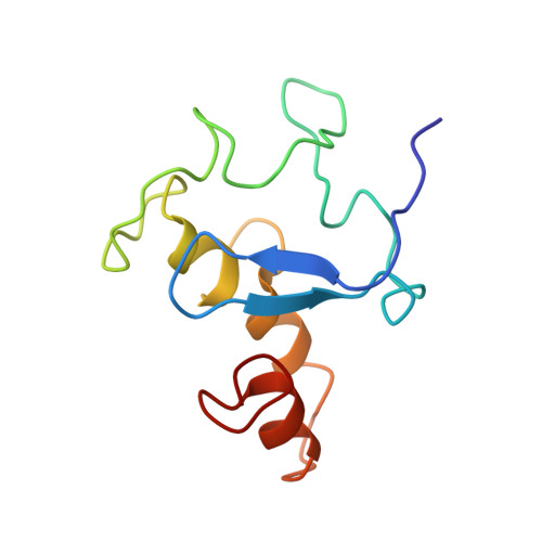
2CDV - PubMed Abstract:
The structure of cytochrome c3 from the sulfate-reducing bacterium Desulfovibrio vulgaris Miyazaki has been successfully refined at 1.8 A resolution. The crystallographic R factor is 0.176 for 9907 significant reflections. The isotropic temperature factors of individual atoms were refined and a total of 47 water molecules located on the difference map were incorporated in the refinement. The four heme groups are closely packed, with adjacent pairs of heme planes being nearly perpendicular to each other. The fifth and the sixth ligands of the heme iron atoms are histidine residues with N epsilon 2-Fe distances ranging from 1.88 A to 2.12 A. The histidine co-ordination to the heme iron is different for each heme group. The heme groups are all highly exposed to solvent, although the actual regions exposed differ among the hemes. The four heme groups are located in different environments, and the heme planes are deformed from planarity. The differences in the heme structures and their environments indicate that the four heme groups are non-equivalent. The chemical as well as the physical properties of cytochrome c3 should be interpreted in terms of the structural non-equivalence of the heme groups. The characteristic secondary structural non-equivalence of the heme groups. The characteristic secondary structures of the polypeptide chain of this molecule are three short alpha-helices, two short beta-strands and ten reverse turns.