2BXX

Crystal structure of the N-terminal domain of IBV coronavirus nucleocapsid. Native crystal form


Experimental Data Snapshot

  • Method: X-RAY DIFFRACTION
  • Resolution: 1.85 Å
  • R-Value Free: 0.270 
  • R-Value Work: 0.230 
  • R-Value Observed: 0.230 

wwPDB Validation   3D Report Full Report


This is version 1.4 of the entry. See complete history


Literature

The Nucleocapsid Protein of Coronavirus Infectious Bronchitis Virus: Crystal Structure of its N-Terminal Domain and Multimerization Properties.

Fan, H.Ooi, A.Tan, Y.W.Wang, S.Fang, S.Liu, D.-X.Lescar, J.

(2005) Structure 13: 1859

  • DOI: https://doi.org/10.1016/j.str.2005.08.021
  • Primary Citation of Related Structures:  
    2BTL, 2BXX

  • PubMed Abstract: 

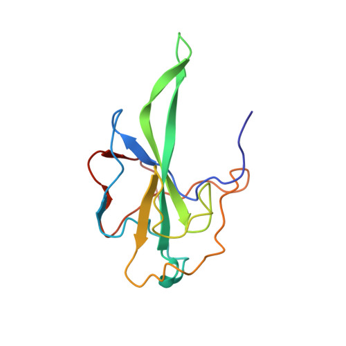
    The coronavirus nucleocapsid (N) protein packages viral genomic RNA into a ribonucleoprotein complex. Interactions between N proteins and RNA are thus crucial for the assembly of infectious virus particles. The 45 kDa recombinant nucleocapsid N protein of coronavirus infectious bronchitis virus (IBV) is highly sensitive to proteolysis. We obtained a stable fragment of 14.7 kDa spanning its N-terminal residues 29-160 (IBV-N29-160). Like the N-terminal RNA binding domain (SARS-N45-181) of the severe acute respiratory syndrome virus (SARS-CoV) N protein, the crystal structure of the IBV-N29-160 fragment at 1.85 A resolution reveals a protein core composed of a five-stranded antiparallel beta sheet with a positively charged beta hairpin extension and a hydrophobic platform that are probably involved in RNA binding. Crosslinking studies demonstrate the formation of dimers, tetramers, and higher multimers of IBV-N. A model for coronavirus shell formation is proposed in which dimerization of the C-terminal domain of IBV-N leads to oligomerization of the IBV-nucleocapsid protein and viral RNA condensation.


  • Organizational Affiliation

    School of Biological Sciences, Nanyang Technological University, 60 Nanyang Drive, Singapore 637551.


Macromolecules
Find similar proteins by:  (by identity cutoff)  |  3D Structure
Entity ID: 1
MoleculeChains Sequence LengthOrganismDetailsImage
NUCLEOCAPSID PROTEIN
A, B
134Infectious bronchitis virusMutation(s): 1 
UniProt
Find proteins for P69596 (Avian infectious bronchitis virus (strain Beaudette))
Explore P69596 
Go to UniProtKB:  P69596
Entity Groups  
Sequence Clusters30% Identity50% Identity70% Identity90% Identity95% Identity100% Identity
UniProt GroupP69596
Sequence Annotations
Expand
  • Reference Sequence
Experimental Data & Validation

Experimental Data

  • Method: X-RAY DIFFRACTION
  • Resolution: 1.85 Å
  • R-Value Free: 0.270 
  • R-Value Work: 0.230 
  • R-Value Observed: 0.230 
  • Space Group: P 1
Unit Cell:
Length ( Å )Angle ( ˚ )
a = 35.422α = 99.2
b = 35.776β = 94.07
c = 55.86γ = 108.96
Software Package:
Software NamePurpose
CNSrefinement

Structure Validation

View Full Validation Report


Entry History 

Deposition Data

Revision History  (Full details and data files)

  • Version 1.0: 2005-12-14
    Type: Initial release
  • Version 1.1: 2011-06-02
    Changes: Version format compliance
  • Version 1.2: 2011-07-13
    Changes: Version format compliance
  • Version 1.3: 2017-06-28
    Changes: Data collection
  • Version 1.4: 2023-12-13
    Changes: Data collection, Database references, Other, Refinement description