1TXQ

Crystal structure of the EB1 C-terminal domain complexed with the CAP-Gly domain of p150Glued


Experimental Data Snapshot

  • Method: X-RAY DIFFRACTION
  • Resolution: 1.80 Å
  • R-Value Free: 0.243 
  • R-Value Work: 0.203 
  • R-Value Observed: 0.203 

wwPDB Validation   3D Report Full Report


This is version 1.3 of the entry. See complete history


Literature

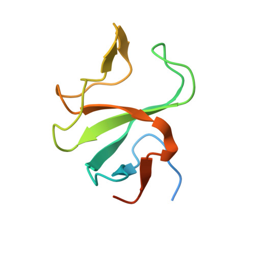
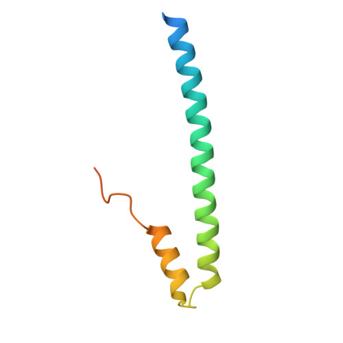
Structural Basis for the Activation of Microtubule Assembly by the EB1 and p150(Glued) Complex

Hayashi, I.Wilde, A.Mal, T.K.Ikura, M.

(2005) Mol Cell 19: 449-460

  • DOI: https://doi.org/10.1016/j.molcel.2005.06.034
  • Primary Citation of Related Structures:  
    1TXQ

  • PubMed Abstract: 

    Plus-end tracking proteins, such as EB1 and the dynein/dynactin complex, regulate microtubule dynamics. These proteins are thought to stabilize microtubules by forming a plus-end complex at microtubule growing ends with ill-defined mechanisms. Here we report the crystal structure of two plus-end complex components, the carboxy-terminal dimerization domain of EB1 and the microtubule binding (CAP-Gly) domain of the dynactin subunit p150Glued. Each molecule of the EB1 dimer contains two helices forming a conserved four-helix bundle, while also providing p150Glued binding sites in its flexible tail region. Combining crystallography, NMR, and mutational analyses, our studies reveal the critical interacting elements of both EB1 and p150Glued, whose mutation alters microtubule polymerization activity. Moreover, removal of the key flexible tail from EB1 activates microtubule assembly by EB1 alone, suggesting that the flexible tail negatively regulates EB1 activity. We, therefore, propose that EB1 possesses an auto-inhibited conformation, which is relieved by p150Glued as an allosteric activator.


  • Organizational Affiliation

    Division of Molecular and Structural Biology, Ontario Cancer Institute and Department of Medical Biophysics, University of Toronto, 610 University Avenue, Toronto, Ontario, M5G 2M9, Canada. ihayashi@uhnres.utoronto.ca


Macromolecules
Find similar proteins by:  (by identity cutoff)  |  3D Structure
Entity ID: 1
MoleculeChains Sequence LengthOrganismDetailsImage
Dynactin 193Homo sapiensMutation(s): 0 
Gene Names: p150Glued
UniProt & NIH Common Fund Data Resources
Find proteins for Q14203 (Homo sapiens)
Explore Q14203 
Go to UniProtKB:  Q14203
PHAROS:  Q14203
GTEx:  ENSG00000204843 
Entity Groups  
Sequence Clusters30% Identity50% Identity70% Identity90% Identity95% Identity100% Identity
UniProt GroupQ14203
Sequence Annotations
Expand
  • Reference Sequence
Find similar proteins by:  (by identity cutoff)  |  3D Structure
Entity ID: 2
MoleculeChains Sequence LengthOrganismDetailsImage
Microtubule-associated protein RP/EB family member 186Homo sapiensMutation(s): 0 
Gene Names: EB1
UniProt & NIH Common Fund Data Resources
Find proteins for Q15691 (Homo sapiens)
Explore Q15691 
Go to UniProtKB:  Q15691
PHAROS:  Q15691
GTEx:  ENSG00000101367 
Entity Groups  
Sequence Clusters30% Identity50% Identity70% Identity90% Identity95% Identity100% Identity
UniProt GroupQ15691
Sequence Annotations
Expand
  • Reference Sequence
Experimental Data & Validation

Experimental Data

  • Method: X-RAY DIFFRACTION
  • Resolution: 1.80 Å
  • R-Value Free: 0.243 
  • R-Value Work: 0.203 
  • R-Value Observed: 0.203 
  • Space Group: C 1 2 1
Unit Cell:
Length ( Å )Angle ( ˚ )
a = 53.085α = 90
b = 80.21β = 108.52
c = 39.239γ = 90
Software Package:
Software NamePurpose
CNSrefinement
HKL-2000data reduction
SCALEPACKdata scaling
SOLVEphasing

Structure Validation

View Full Validation Report


Entry History 

Deposition Data

Revision History  (Full details and data files)

  • Version 1.0: 2005-09-13
    Type: Initial release
  • Version 1.1: 2008-04-30
    Changes: Version format compliance
  • Version 1.2: 2011-07-13
    Changes: Version format compliance
  • Version 1.3: 2024-03-13
    Changes: Data collection, Database references