1EFH

CRYSTAL STRUCTURE OF THE HUMAN HYDROXYSTEROID SULFOTRANSFERASE IN THE PRESENCE OF PAP


Experimental Data Snapshot

  • Method: X-RAY DIFFRACTION
  • Resolution: 2.40 Å
  • R-Value Free: 0.239 
  • R-Value Work: 0.202 
  • R-Value Observed: 0.204 

wwPDB Validation   3D Report Full Report


Ligand Structure Quality Assessment 


This is version 1.4 of the entry. See complete history


Literature

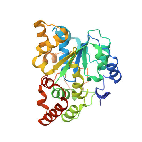
Crystal structure of SULT2A3, human hydroxysteroid sulfotransferase.

Pedersen, L.C.Petrotchenko, E.V.Negishi, M.

(2000) FEBS Lett 475: 61-64

  • DOI: https://doi.org/10.1016/s0014-5793(00)01479-4
  • Primary Citation of Related Structures:  
    1EFH

  • PubMed Abstract: 

    The crystal structure of SULT2A3 human hydroxysteroid sulfotransferase has been solved at 2.4 A resolution in the presence of 3'-phosphoadenosine 5'-phosphate (PAP). The overall structure is similar to those of SULT1 enzymes such as estrogen sulfotransferase and the PAP binding site is conserved, however, significant differences exist in the positions of loops Pro14-Ser20, Glu79-Ile82 and Tyr234-Gln244 in the substrate binding pocket. Moreover, protein interaction in the crystal structure has revealed a possible dimer-directed conformational alteration that may regulate the SULT activity.


  • Organizational Affiliation

    Pharmacogenetics Section, Laboratory of Reproductive and Developmental Toxicology, National Institute of Environmental Health Sciences, National Institutes of Health, Research Triangle Park, NC 27709, USA.


Macromolecules
Find similar proteins by:  (by identity cutoff)  |  3D Structure
Entity ID: 1
MoleculeChains Sequence LengthOrganismDetailsImage
HYDROXYSTEROID SULFOTRANSFERASE
A, B
292Homo sapiensMutation(s): 0 
Gene Names: CDNA LIBRARY
EC: 2.8.2.2
UniProt & NIH Common Fund Data Resources
Find proteins for Q06520 (Homo sapiens)
Explore Q06520 
Go to UniProtKB:  Q06520
PHAROS:  Q06520
GTEx:  ENSG00000105398 
Entity Groups  
Sequence Clusters30% Identity50% Identity70% Identity90% Identity95% Identity100% Identity
UniProt GroupQ06520
Sequence Annotations
Expand
  • Reference Sequence
Small Molecules
Ligands 1 Unique
IDChains Name / Formula / InChI Key2D Diagram3D Interactions
A3P
Query on A3P

Download Ideal Coordinates CCD File 
C [auth A],
D [auth B]
ADENOSINE-3'-5'-DIPHOSPHATE
C10 H15 N5 O10 P2
WHTCPDAXWFLDIH-KQYNXXCUSA-N
Experimental Data & Validation

Experimental Data

  • Method: X-RAY DIFFRACTION
  • Resolution: 2.40 Å
  • R-Value Free: 0.239 
  • R-Value Work: 0.202 
  • R-Value Observed: 0.204 
  • Space Group: P 21 21 21
Unit Cell:
Length ( Å )Angle ( ˚ )
a = 73.19α = 90
b = 96.82β = 90
c = 127.38γ = 90
Software Package:
Software NamePurpose
AMoREphasing
CNSrefinement
DENZOdata reduction
SCALEPACKdata scaling

Structure Validation

View Full Validation Report


Ligand Structure Quality Assessment 


Entry History 

Deposition Data

Revision History  (Full details and data files)

  • Version 1.0: 2000-09-06
    Type: Initial release
  • Version 1.1: 2008-04-27
    Changes: Version format compliance
  • Version 1.2: 2011-07-13
    Changes: Version format compliance
  • Version 1.3: 2018-06-06
    Changes: Data collection, Derived calculations
  • Version 1.4: 2024-02-07
    Changes: Data collection, Database references, Derived calculations