1ZET

X-ray data do not support hoogsteen base-pairing during replication by human polymerase iota


Experimental Data Snapshot

  • Method: X-RAY DIFFRACTION
  • Resolution: 2.30 Å
  • R-Value Free: 0.288 
  • R-Value Work: 0.261 
  • R-Value Observed: 0.261 

wwPDB Validation   3D Report Full Report


Ligand Structure Quality Assessment 


This is version 1.4 of the entry. See complete history

Re-refinement Note

This entry reflects an alternative modeling of the original data in: 1T3N


Literature

DNA polymerases: Hoogsteen base-pairing in DNA replication?

Wang, J.

(2005) Nature 437: E6-7; discussion E7

  • DOI: https://doi.org/10.1038/nature04199
  • Primary Citation of Related Structures:  
    1ZET

  • PubMed Abstract: 

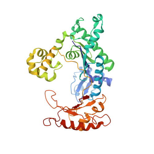
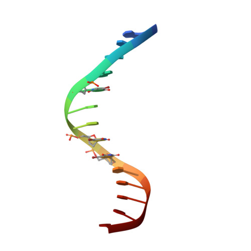
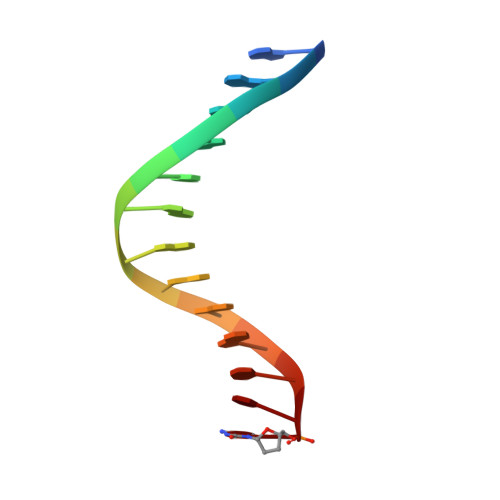
    Human polymerase-iota belongs to the error-prone Y family of polymerases, which frequently incorporate incorrect nucleotides during DNA replication but can efficiently bypass DNA lesions. On the basis of X-ray diffraction data, Nair et al. propose that Hoogsteen base-pairing is adopted by DNA during its replication by this enzyme. Here I re-examine their X-ray data and find that the electron density is very weak for a Hoogsteen base pair formed between a template adenine deoxyribonucleotide in the syn conformation and a deoxythymidine 5'-triphosphate (dTTP), and that the fit is better for a normal Watson-Crick base pair. As a guanine-cytosine (G-C) base pair has no potential to form a Hoogsteen base pair at physiological pH, Hoogsteen base-pairing is unlikely to be used in replication by this polymerase.


  • Organizational Affiliation

    Center for Structural Biology, Department of Molecular Biophysics and Biochemistry, Yale University, New Haven, Connecticut 06520, USA wang@csb.yale.edu.


Macromolecules

Find similar proteins by:  (by identity cutoff)  |  3D Structure
Entity ID: 3
MoleculeChains Sequence LengthOrganismDetailsImage
Polymerase (DNA directed) iotaC [auth A]388Homo sapiensMutation(s): 0 
Gene Names: POLI
UniProt & NIH Common Fund Data Resources
Find proteins for Q9UNA4 (Homo sapiens)
Explore Q9UNA4 
Go to UniProtKB:  Q9UNA4
PHAROS:  Q9UNA4
GTEx:  ENSG00000101751 
Entity Groups  
Sequence Clusters30% Identity50% Identity70% Identity90% Identity95% Identity100% Identity
UniProt GroupQ9UNA4
Sequence Annotations
Expand
  • Reference Sequence

Find similar nucleic acids by:  Sequence   |   3D Structure  

Entity ID: 1
MoleculeChains LengthOrganismImage
5'-D(P*AP*GP*GP*GP*(BRU)P*CP*CP*(BRU)P*(BRU)P*CP*CP*CP*CP*C)-3'A [auth T]14N/A
Sequence Annotations
Expand
  • Reference Sequence

Find similar nucleic acids by:  Sequence   |   3D Structure  

Entity ID: 2
MoleculeChains LengthOrganismImage
5'-D(*GP*GP*GP*GP*GP*AP*AP*GP*GP*AP*CP*CP*(DOC))-3'B [auth P]13N/A
Sequence Annotations
Expand
  • Reference Sequence
Small Molecules
Ligands 2 Unique
IDChains Name / Formula / InChI Key2D Diagram3D Interactions
TTP
Query on TTP

Download Ideal Coordinates CCD File 
E [auth A]THYMIDINE-5'-TRIPHOSPHATE
C10 H17 N2 O14 P3
NHVNXKFIZYSCEB-XLPZGREQSA-N
MG
Query on MG

Download Ideal Coordinates CCD File 
D [auth A]MAGNESIUM ION
Mg
JLVVSXFLKOJNIY-UHFFFAOYSA-N
Experimental Data & Validation

Experimental Data

  • Method: X-RAY DIFFRACTION
  • Resolution: 2.30 Å
  • R-Value Free: 0.288 
  • R-Value Work: 0.261 
  • R-Value Observed: 0.261 
  • Space Group: P 65 2 2
Unit Cell:
Length ( Å )Angle ( ˚ )
a = 98.831α = 90
b = 98.831β = 90
c = 202.755γ = 120
Software Package:
Software NamePurpose
CNSrefinement

Structure Validation

View Full Validation Report


Ligand Structure Quality Assessment 


Entry History 

Deposition Data

  • Released Date: 2005-07-19 
  • Deposition Author(s): Wang, J.

Revision History  (Full details and data files)

  • Version 1.0: 2005-07-19
    Type: Initial release
  • Version 1.1: 2008-04-30
    Changes: Version format compliance
  • Version 1.2: 2011-07-13
    Changes: Version format compliance
  • Version 1.3: 2011-09-28
    Changes: Other
  • Version 1.4: 2024-02-14
    Changes: Data collection, Database references, Derived calculations