Structural basis of spectral shifts in the yellow-emission variants of green fluorescent protein.
Wachter, R.M., Elsliger, M.A., Kallio, K., Hanson, G.T., Remington, S.J.(1998) Structure 6: 1267-1277
- PubMed: 9782051
- DOI: https://doi.org/10.1016/s0969-2126(98)00127-0
- Primary Citation of Related Structures:
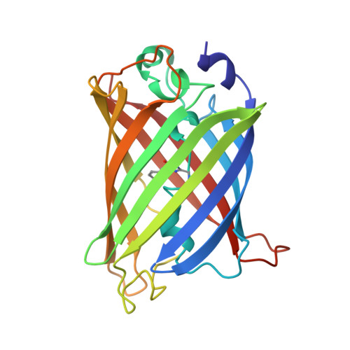
1YFP, 2YFP - PubMed Abstract:
Because of its ability to spontaneously generate its own fluorophore, the green fluorescent protein (GFP) from the jellyfish Aequorea victoria is used extensively as a fluorescent marker in molecular and cell biology. The yellow fluorescent proteins (YFPs) have the longest wavelength emissions of all GFP variants examined to date. This shift in the spectrum is the result of a T203Y substitution (single-letter amino acid code), a mutation rationally designed on the basis of the X-ray structure of GFP S65T. We have determined the crystal structures of YFP T203Y/S65G/V68L/S72A and YFP H148G to 2.5 and 2.6 A resolution, respectively. Both structures show clear electron density for nearly coplanar pi-pi stacking between Tyr203 and the chromophore. The chromophore has been displaced by nearly 1 A in comparison to other available structures. Although the H148G mutation results in the generation of a solvent channel to the chromophore cavity, intense fluorescence is maintained. The chromophore in the intact protein can be titrated, and the two variants have pKa values of 7.0 (YFP) and 8.0 (YFP H148G). The observed red shift of the T203Y YFP variant is proposed to be mainly due to the additional polarizability of the pi-stacked Tyr203. The altered location of the chromophore suggests that the exact positions of nearby residues are not crucial for the chemistry of chromophore formation. The YFPs significantly extend the pH range over which GFPs may be employed as pH indicators in live cells.
Organizational Affiliation:
Institute of Molecular Biology, Department of Physics, University of Oregon, Eugene, Oregon, 97403 USA.