1TN6

Protein Farnesyltransferase Complexed with a Rap2a Peptide Substrate and a FPP Analog at 1.8A Resolution


Experimental Data Snapshot

  • Method: X-RAY DIFFRACTION
  • Resolution: 1.80 Å
  • R-Value Free: 0.199 
  • R-Value Work: 0.179 
  • R-Value Observed: 0.179 

wwPDB Validation   3D Report Full Report


Ligand Structure Quality Assessment 


This is version 2.1 of the entry. See complete history


Literature

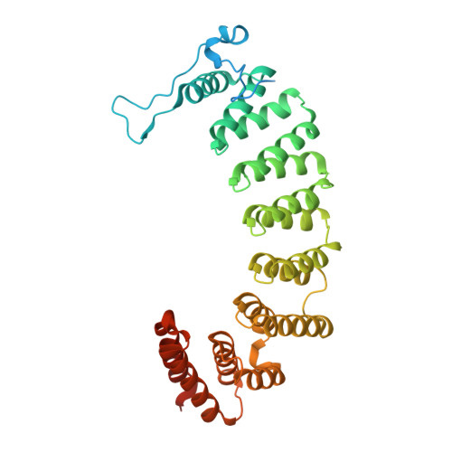
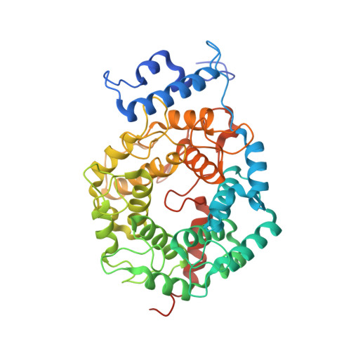
Crystallographic analysis of CaaX prenyltransferases complexed with substrates defines rules of protein substrate selectivity

Reid, T.S.Terry, K.L.Casey, P.J.Beese, L.S.

(2004) J Mol Biol 343: 417-433

  • DOI: https://doi.org/10.1016/j.jmb.2004.08.056
  • Primary Citation of Related Structures:  
    1TN6, 1TN7, 1TN8, 1TNB, 1TNO, 1TNU, 1TNY, 1TNZ

  • PubMed Abstract: 

    Post-translational modifications are essential for the proper function of many proteins in the cell. The attachment of an isoprenoid lipid (a process termed prenylation) by protein farnesyltransferase (FTase) or geranylgeranyltransferase type I (GGTase-I) is essential for the function of many signal transduction proteins involved in growth, differentiation, and oncogenesis. FTase and GGTase-I (also called the CaaX prenyltransferases) recognize protein substrates with a C-terminal tetrapeptide recognition motif called the Ca1a2X box. These enzymes possess distinct but overlapping protein substrate specificity that is determined primarily by the sequence identity of the Ca1a2X motif. To determine how the identity of the Ca1a2X motif residues and sequence upstream of this motif affect substrate binding, we have solved crystal structures of FTase and GGTase-I complexed with a total of eight cognate and cross-reactive substrate peptides, including those derived from the C termini of the oncoproteins K-Ras4B, H-Ras and TC21. These structures suggest that all peptide substrates adopt a common binding mode in the FTase and GGTase-I active site. Unexpectedly, while the X residue of the Ca1a2X motif binds in the same location for all GGTase-I substrates, the X residue of FTase substrates can bind in one of two different sites. Together, these structures outline a series of rules that govern substrate peptide selectivity; these rules were utilized to classify known protein substrates of CaaX prenyltransferases and to generate a list of hypothetical substrates within the human genome.


  • Organizational Affiliation

    Department of Biochemistry, Duke University Medical Center, Durham, NC 27710, USA.


Macromolecules
Find similar proteins by:  (by identity cutoff)  |  3D Structure
Entity ID: 1
MoleculeChains Sequence LengthOrganismDetailsImage
Protein farnesyltransferase alpha subunit382Homo sapiensMutation(s): 0 
Gene Names: FNTA
EC: 2.5.1.58
UniProt & NIH Common Fund Data Resources
Find proteins for P49354 (Homo sapiens)
Explore P49354 
Go to UniProtKB:  P49354
PHAROS:  P49354
GTEx:  ENSG00000168522 
Entity Groups  
Sequence Clusters30% Identity50% Identity70% Identity90% Identity95% Identity100% Identity
UniProt GroupP49354
Sequence Annotations
Expand
  • Reference Sequence
Find similar proteins by:  (by identity cutoff)  |  3D Structure
Entity ID: 2
MoleculeChains Sequence LengthOrganismDetailsImage
Protein farnesyltransferase beta subunit437Homo sapiensMutation(s): 0 
Gene Names: FNTB
EC: 2.5.1.58
UniProt & NIH Common Fund Data Resources
Find proteins for P49356 (Homo sapiens)
Explore P49356 
Go to UniProtKB:  P49356
PHAROS:  P49356
GTEx:  ENSG00000257365 
Entity Groups  
Sequence Clusters30% Identity50% Identity70% Identity90% Identity95% Identity100% Identity
UniProt GroupP49356
Sequence Annotations
Expand
  • Reference Sequence

Find similar proteins by:  Sequence   |   3D Structure  

Entity ID: 3
MoleculeChains Sequence LengthOrganismDetailsImage
peptide derived from the C-terminus of Rap2a11N/AMutation(s): 0 
Entity Groups  
Sequence Clusters30% Identity50% Identity70% Identity90% Identity95% Identity100% Identity
Sequence Annotations
Expand
  • Reference Sequence
Oligosaccharides

Help

Entity ID: 4
MoleculeChains Length2D Diagram Glycosylation3D Interactions
beta-D-fructofuranose-(2-1)-alpha-D-glucopyranose
D
2N/A
Glycosylation Resources
GlyTouCan:  G05551OP
GlyCosmos:  G05551OP
Biologically Interesting Molecules (External Reference) 1 Unique
Experimental Data & Validation

Experimental Data

  • Method: X-RAY DIFFRACTION
  • Resolution: 1.80 Å
  • R-Value Free: 0.199 
  • R-Value Work: 0.179 
  • R-Value Observed: 0.179 
  • Space Group: P 61
Unit Cell:
Length ( Å )Angle ( ˚ )
a = 178.474α = 90
b = 178.474β = 90
c = 64.697γ = 120
Software Package:
Software NamePurpose
CNSrefinement
DENZOdata reduction
SCALEPACKdata scaling
CNSphasing

Structure Validation

View Full Validation Report


Ligand Structure Quality Assessment 


Entry History 

Deposition Data

Revision History  (Full details and data files)

  • Version 1.0: 2004-11-02
    Type: Initial release
  • Version 1.1: 2008-04-30
    Changes: Version format compliance
  • Version 1.2: 2011-07-13
    Changes: Version format compliance
  • Version 2.0: 2020-07-29
    Type: Remediation
    Reason: Carbohydrate remediation
    Changes: Atomic model, Data collection, Database references, Derived calculations, Non-polymer description, Structure summary
  • Version 2.1: 2023-08-23
    Changes: Data collection, Database references, Refinement description, Structure summary