1THQ

Crystal Structure of Outer Membrane Enzyme PagP


Experimental Data Snapshot

  • Method: X-RAY DIFFRACTION
  • Resolution: 1.90 Å
  • R-Value Free: 0.255 
  • R-Value Work: 0.216 
  • R-Value Observed: 0.218 

wwPDB Validation   3D Report Full Report


Ligand Structure Quality Assessment 


This is version 1.3 of the entry. See complete history


Literature

A hydrocarbon ruler measures palmitate in the enzymatic acylation of endotoxin.

Ahn, V.E.Lo, E.I.Engel, C.K.Chen, L.Hwang, P.M.Kay, L.E.Bishop, R.E.Prive, G.G.

(2004) EMBO J 23: 2931-2941

  • DOI: https://doi.org/10.1038/sj.emboj.7600320
  • Primary Citation of Related Structures:  
    1THQ

  • PubMed Abstract: 

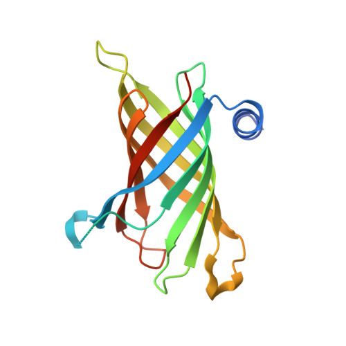
    The ability of enzymes to distinguish between fatty acyl groups can involve molecular measuring devices termed hydrocarbon rulers, but the molecular basis for acyl-chain recognition in any membrane-bound enzyme remains to be defined. PagP is an outer membrane acyltransferase that helps pathogenic bacteria to evade the host immune response by transferring a palmitate chain from a phospholipid to lipid A (endotoxin). PagP can distinguish lipid acyl chains that differ by a single methylene unit, indicating that the enzyme possesses a remarkably precise hydrocarbon ruler. We present the 1.9 A crystal structure of PagP, an eight-stranded beta-barrel with an unexpected interior hydrophobic pocket that is occupied by a single detergent molecule. The buried detergent is oriented normal to the presumed plane of the membrane, whereas the PagP beta-barrel axis is tilted by approximately 25 degrees. Acyl group specificity is modulated by mutation of Gly88 lining the bottom of the hydrophobic pocket, thus confirming the hydrocarbon ruler mechanism for palmitate recognition. A striking structural similarity between PagP and the lipocalins suggests an evolutionary link between these proteins.


  • Organizational Affiliation

    Department of Medical Biophysics, University of Toronto, Ontario, Canada.


Macromolecules
Find similar proteins by:  (by identity cutoff)  |  3D Structure
Entity ID: 1
MoleculeChains Sequence LengthOrganismDetailsImage
CrcA protein170Escherichia coliMutation(s): 0 
Gene Names: CRCAB0622
Membrane Entity: Yes 
UniProt
Find proteins for P37001 (Escherichia coli (strain K12))
Explore P37001 
Go to UniProtKB:  P37001
Entity Groups  
Sequence Clusters30% Identity50% Identity70% Identity90% Identity95% Identity100% Identity
UniProt GroupP37001
Sequence Annotations
Expand
  • Reference Sequence
Small Molecules
Ligands 3 Unique
IDChains Name / Formula / InChI Key2D Diagram3D Interactions
LDA
Query on LDA

Download Ideal Coordinates CCD File 
G [auth A],
H [auth A],
I [auth A],
J [auth A],
K [auth A]
LAURYL DIMETHYLAMINE-N-OXIDE
C14 H31 N O
SYELZBGXAIXKHU-UHFFFAOYSA-N
MPD
Query on MPD

Download Ideal Coordinates CCD File 
L [auth A],
M [auth A]
(4S)-2-METHYL-2,4-PENTANEDIOL
C6 H14 O2
SVTBMSDMJJWYQN-YFKPBYRVSA-N
ACT
Query on ACT

Download Ideal Coordinates CCD File 
B [auth A],
C [auth A],
D [auth A],
E [auth A],
F [auth A]
ACETATE ION
C2 H3 O2
QTBSBXVTEAMEQO-UHFFFAOYSA-M
Experimental Data & Validation

Experimental Data

  • Method: X-RAY DIFFRACTION
  • Resolution: 1.90 Å
  • R-Value Free: 0.255 
  • R-Value Work: 0.216 
  • R-Value Observed: 0.218 
  • Space Group: P 41 2 2
Unit Cell:
Length ( Å )Angle ( ˚ )
a = 50.67α = 90
b = 50.67β = 90
c = 158.48γ = 90
Software Package:
Software NamePurpose
REFMACrefinement
DENZOdata reduction
SCALEPACKdata scaling
SOLVEphasing

Structure Validation

View Full Validation Report


Ligand Structure Quality Assessment 


Entry History 

Deposition Data

Revision History  (Full details and data files)

  • Version 1.0: 2004-08-10
    Type: Initial release
  • Version 1.1: 2008-04-30
    Changes: Version format compliance
  • Version 1.2: 2011-07-13
    Changes: Advisory, Version format compliance
  • Version 1.3: 2024-02-14
    Changes: Data collection, Database references, Derived calculations