Structural test of the parameterized-backbone method for protein design
Plecs, J.J., Harbury, P.B., Kim, P.S., Alber, T.(2004) J Mol Biol 342: 289-297
- PubMed: 15313624
- DOI: https://doi.org/10.1016/j.jmb.2004.06.051
- Primary Citation of Related Structures:
1TGG - PubMed Abstract:
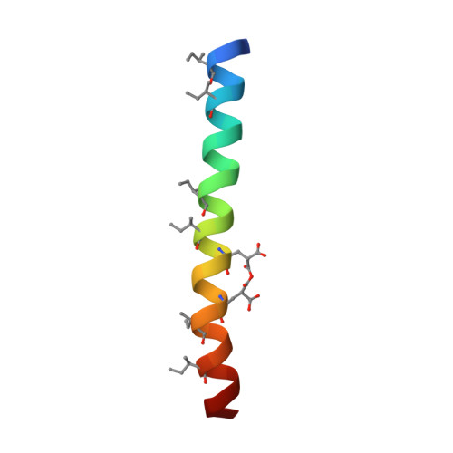
Designing new protein folds requires a method for simultaneously optimizing the conformation of the backbone and the side-chains. One approach to this problem is the use of a parameterized backbone, which allows the systematic exploration of families of structures. We report the crystal structure of RH3, a right-handed, three-helix coiled coil that was designed using a parameterized backbone and detailed modeling of core packing. This crystal structure was determined using another rationally designed feature, a metal-binding site that permitted experimental phasing of the X-ray data. RH3 adopted the intended fold, which has not been observed previously in biological proteins. Unanticipated structural asymmetry in the trimer was a principal source of variation within the RH3 structure. The sequence of RH3 differs from that of a previously characterized right-handed tetramer, RH4, at only one position in each 11 amino acid sequence repeat. This close similarity indicates that the design method is sensitive to the core packing interactions that specify the protein structure. Comparison of the structures of RH3 and RH4 indicates that both steric overlap and cavity formation provide strong driving forces for oligomer specificity.
Organizational Affiliation:
Department of Physics, University of California, Berkeley, 94720, USA.