1S1W

Crystal structure of V106A mutant HIV-1 reverse transcriptase in complex with UC-781


Experimental Data Snapshot

  • Method: X-RAY DIFFRACTION
  • Resolution: 2.70 Å
  • R-Value Free: 0.280 
  • R-Value Work: 0.206 
  • R-Value Observed: 0.198 

wwPDB Validation   3D Report Full Report


This is version 1.3 of the entry. See complete history


Literature

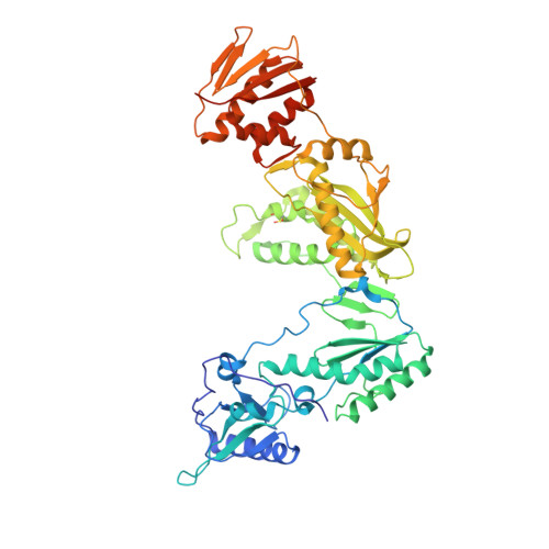
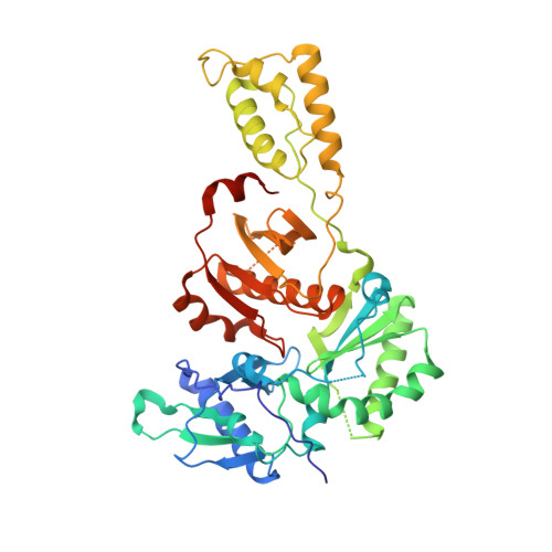
Crystal structures of HIV-1 reverse transcriptases mutated at codons 100, 106 and 108 and mechanisms of resistance to non-nucleoside inhibitors

Ren, J.Nichols, C.E.Chamberlain, P.P.Weaver, K.L.Short, S.A.Stammers, D.K.

(2004) J Mol Biol 336: 569-578

  • DOI: https://doi.org/10.1016/j.jmb.2003.12.055
  • Primary Citation of Related Structures:  
    1S1T, 1S1U, 1S1V, 1S1W, 1S1X

  • PubMed Abstract: 

    Leu100Ile, Val106Ala and Val108Ile are mutations in HIV-1 reverse transcriptase (RT) that are observed in the clinic and give rise to resistance to certain non-nucleoside inhibitors (NNRTIs) including the first-generation drug nevirapine. In order to investigate structural mechanisms of resistance for different NNRTI classes we have determined six crystal structures of mutant RT-inhibitor complexes. Val108 does not have direct contact with nevirapine in wild-type RT and in the RT(Val108Ile) complex the biggest change observed is at the distally positioned Tyr181 which is > 8 A from the mutation site. Thus in contrast to most NNRTI resistance mutations RT(Val108Ile) appears to act via an indirect mechanism which in this case is through alterations of the ring stacking interactions of the drug particularly with Tyr181. Shifts in side-chain and inhibitor positions compared to wild-type RT are observed in complexes of nevirapine and the second-generation NNRTI UC-781 with RT(Leu100Ile) and RT(Val106Ala), leading to perturbations in inhibitor contacts with Tyr181 and Tyr188. Such perturbations are likely to be a factor contributing to the greater loss of binding for nevirapine compared to UC-781 as, in the former case, a larger proportion of binding energy is derived from aromatic ring stacking of the inhibitor with the tyrosine side-chains. The differing resistance profiles of first and second generation NNRTIs for other drug resistance mutations in RT may also be in part due to this indirect mechanism.


  • Organizational Affiliation

    Division of Structural Biology, The Wellcome Trust Centre for Human Genetics, Henry Wellcome Building for Genomic Medicine, University of Oxford Roosevelt Drive, Oxford OX3 7BN, UK.


Macromolecules
Find similar proteins by:  (by identity cutoff)  |  3D Structure
Entity ID: 1
MoleculeChains Sequence LengthOrganismDetailsImage
Reverse transcriptase560Human immunodeficiency virus 1Mutation(s): 2 
Gene Names: POL
EC: 2.7.7.49
UniProt
Find proteins for P04585 (Human immunodeficiency virus type 1 group M subtype B (isolate HXB2))
Explore P04585 
Go to UniProtKB:  P04585
Entity Groups  
Sequence Clusters30% Identity50% Identity70% Identity90% Identity95% Identity100% Identity
UniProt GroupP04585
Sequence Annotations
Expand
  • Reference Sequence
Find similar proteins by:  (by identity cutoff)  |  3D Structure
Entity ID: 2
MoleculeChains Sequence LengthOrganismDetailsImage
Reverse transcriptase440Human immunodeficiency virus 1Mutation(s): 1 
Gene Names: POL
EC: 2.7.7.49
UniProt
Find proteins for P04585 (Human immunodeficiency virus type 1 group M subtype B (isolate HXB2))
Explore P04585 
Go to UniProtKB:  P04585
Entity Groups  
Sequence Clusters30% Identity50% Identity70% Identity90% Identity95% Identity100% Identity
UniProt GroupP04585
Sequence Annotations
Expand
  • Reference Sequence
Small Molecules
Ligands 1 Unique
IDChains Name / Formula / InChI Key2D Diagram3D Interactions
UC1
Query on UC1

Download Ideal Coordinates CCD File 
C [auth A]2-METHYL-FURAN-3-CARBOTHIOIC ACID [4-CHLORO-3-(3-METHYL-BUT-2-ENYLOXY)-PHENYL]-AMIDE
C17 H18 Cl N O2 S
YZHIXLCGPOTQNB-UHFFFAOYSA-N
Modified Residues  1 Unique
IDChains TypeFormula2D DiagramParent
CSD
Query on CSD
A
L-PEPTIDE LINKINGC3 H7 N O4 SCYS
Binding Affinity Annotations 
IDSourceBinding Affinity
UC1 BindingDB:  1S1W IC50: min: 20.8, max: 226 (nM) from 3 assay(s)
EC50: min: 9, max: 24 (nM) from 2 assay(s)
Experimental Data & Validation

Experimental Data

  • Method: X-RAY DIFFRACTION
  • Resolution: 2.70 Å
  • R-Value Free: 0.280 
  • R-Value Work: 0.206 
  • R-Value Observed: 0.198 
  • Space Group: P 21 21 21
Unit Cell:
Length ( Å )Angle ( ˚ )
a = 137.54α = 90
b = 114.69β = 90
c = 65.26γ = 90
Software Package:
Software NamePurpose
CNSrefinement
DENZOdata reduction
SCALEPACKdata scaling
CNSphasing

Structure Validation

View Full Validation Report


Entry History 

Deposition Data

Revision History  (Full details and data files)

  • Version 1.0: 2004-06-29
    Type: Initial release
  • Version 1.1: 2008-04-29
    Changes: Version format compliance
  • Version 1.2: 2011-07-13
    Changes: Version format compliance
  • Version 1.3: 2021-11-10
    Changes: Database references, Derived calculations