1S1S

Crystal Structure of ZipA in complex with indoloquinolizin 10b


Experimental Data Snapshot

  • Method: X-RAY DIFFRACTION
  • Resolution: 2.10 Å
  • R-Value Free: 0.280 
  • R-Value Work: 0.226 
  • R-Value Observed: 0.226 

wwPDB Validation   3D Report Full Report


This is version 1.3 of the entry. See complete history


Literature

Design and synthesis of indolo[2,3-a]quinolizin-7-one inhibitors of the ZipA-FtsZ interaction

JENNINGS, L.D.FOREMAN, K.W.RUSH III, T.S.TSAO, D.H.MOSYAK, L.LI, Y.SUKHDEO, M.N.DING, W.DUSHIN, E.G.KENNY, C.H.MOGHAZEH, S.L.PETERSEN, P.J.RUZIN, A.V.TUCKMAN, M.SUTHERLAND, A.G.

(2004) Bioorg Med Chem Lett 14: 1427-1431

  • DOI: https://doi.org/10.1016/j.bmcl.2004.01.028
  • Primary Citation of Related Structures:  
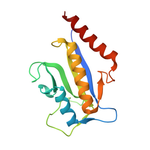
    1S1J, 1S1S

  • PubMed Abstract: 

    The binding of FtsZ to ZipA is a potential target for antibacterial therapy. Based on a small molecule inhibitor of the ZipA-FtsZ interaction, a parallel synthesis of small molecules was initiated which targeted a key region of ZipA involved in FtsZ binding. The X-ray crystal structure of one of these molecules complexed with ZipA was solved. The structure revealed an unexpected binding mode, facilitated by desolvation of a loosely bound surface water.


  • Organizational Affiliation

    Wyeth Research, Department of Medicinal Chemistry, 401N. Middletown Rd, Pearl River, NY 10965, USA. jenninl@wyeth.com


Macromolecules
Find similar proteins by:  (by identity cutoff)  |  3D Structure
Entity ID: 1
MoleculeChains Sequence LengthOrganismDetailsImage
Cell division protein zipA
A, B
144Escherichia coliMutation(s): 0 
Gene Names: ZIPAB2412
UniProt
Find proteins for P77173 (Escherichia coli (strain K12))
Explore P77173 
Go to UniProtKB:  P77173
Entity Groups  
Sequence Clusters30% Identity50% Identity70% Identity90% Identity95% Identity100% Identity
UniProt GroupP77173
Sequence Annotations
Expand
  • Reference Sequence
Small Molecules
Ligands 1 Unique
IDChains Name / Formula / InChI Key2D Diagram3D Interactions
WAC
Query on WAC

Download Ideal Coordinates CCD File 
C [auth B]N-{3-[(12bS)-7-oxo-1,3,4,6,7,12b-hexahydroindolo[2,3-a]quinolizin-12(2H)-yl]propyl}propane-2-sulfonamide
C21 H29 N3 O3 S
LICJTIDRHJECTD-SFHVURJKSA-N
Experimental Data & Validation

Experimental Data

  • Method: X-RAY DIFFRACTION
  • Resolution: 2.10 Å
  • R-Value Free: 0.280 
  • R-Value Work: 0.226 
  • R-Value Observed: 0.226 
  • Space Group: P 1 21 1
Unit Cell:
Length ( Å )Angle ( ˚ )
a = 52.446α = 90
b = 38.847β = 106.28
c = 71.515γ = 90
Software Package:
Software NamePurpose
CNSrefinement
AMoREphasing

Structure Validation

View Full Validation Report


Entry History 

Revision History  (Full details and data files)

  • Version 1.0: 2005-01-18
    Type: Initial release
  • Version 1.1: 2008-04-29
    Changes: Version format compliance
  • Version 1.2: 2011-07-13
    Changes: Version format compliance
  • Version 1.3: 2024-02-14
    Changes: Data collection, Database references, Derived calculations