1R5L

Crystal Structure of Human Alpha-Tocopherol Transfer Protein Bound to its Ligand


Experimental Data Snapshot

  • Method: X-RAY DIFFRACTION
  • Resolution: 1.50 Å
  • R-Value Free: 0.213 
  • R-Value Work: 0.187 
  • R-Value Observed: 0.187 

wwPDB Validation   3D Report Full Report


Ligand Structure Quality Assessment 


This is version 1.2 of the entry. See complete history


Literature

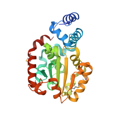
Crystal structure of human alpha-tocopherol transfer protein bound to its ligand: Implications for ataxia with vitamin E deficiency

Min, K.C.Kovall, R.A.Hendrickson, W.A.

(2003) Proc Natl Acad Sci U S A 100: 14713-14718

  • DOI: https://doi.org/10.1073/pnas.2136684100
  • Primary Citation of Related Structures:  
    1R5L

  • PubMed Abstract: 

    Human alpha-tocopherol (alpha-T) transfer protein (ATTP) plays a central role in vitamin E homeostasis, preventing degradation of alpha-T by routing this lipophilic molecule for secretion by hepatocytes. Mutations in the gene encoding ATTP have been shown to cause a severe deficiency in alpha-T, which results in a progressive neurodegenerative spinocerebellar ataxia, known as ataxia with vitamin E deficiency (AVED). We have determined the high-resolution crystal structure of human ATTP with (2R,4'R,8'R)-alpha-T in the binding pocket. Surprisingly, the ligand is sequestered deep in the hydrophobic core of the protein, implicating a large structural rearrangement for the entry and release of alpha-T. A comparison to the structure of a related protein, Sec14p, crystallized without a bona fide ligand, shows a possibly relevant open conformation for this family of proteins. Furthermore, of the known mutations that cause AVED, one mutation, L183P, is located directly in the binding pocket. Finally, three mutations associated with AVED involve arginine residues that are grouped together on the surface of ATTP. We propose that this positively charged surface may serve to orient an interacting protein, which might function to regulate the release of alpha-T through an induced change in conformation of ATTP.


  • Organizational Affiliation

    Howard Hughes Medical Institute and Department of Neurology, Columbia University, New York, NY 10032, USA.


Macromolecules
Find similar proteins by:  (by identity cutoff)  |  3D Structure
Entity ID: 1
MoleculeChains Sequence LengthOrganismDetailsImage
PROTEIN (Alpha-tocopherol transfer protein)262Homo sapiensMutation(s): 4 
Gene Names: TTPA OR TPP1
UniProt & NIH Common Fund Data Resources
Find proteins for P49638 (Homo sapiens)
Explore P49638 
Go to UniProtKB:  P49638
PHAROS:  P49638
GTEx:  ENSG00000137561 
Entity Groups  
Sequence Clusters30% Identity50% Identity70% Identity90% Identity95% Identity100% Identity
UniProt GroupP49638
Sequence Annotations
Expand
  • Reference Sequence
Small Molecules
Ligands 1 Unique
IDChains Name / Formula / InChI Key2D Diagram3D Interactions
VIV
Query on VIV

Download Ideal Coordinates CCD File 
B [auth A](2R)-2,5,7,8-TETRAMETHYL-2-[(4R,8R)-4,8,12-TRIMETHYLTRIDECYL]CHROMAN-6-OL
C29 H50 O2
GVJHHUAWPYXKBD-IEOSBIPESA-N
Modified Residues  1 Unique
IDChains TypeFormula2D DiagramParent
MSE
Query on MSE
A
L-PEPTIDE LINKINGC5 H11 N O2 SeMET
Binding Affinity Annotations 
IDSourceBinding Affinity
VIV BindingDB:  1R5L Kd: 2.4 (nM) from 1 assay(s)
Experimental Data & Validation

Experimental Data

  • Method: X-RAY DIFFRACTION
  • Resolution: 1.50 Å
  • R-Value Free: 0.213 
  • R-Value Work: 0.187 
  • R-Value Observed: 0.187 
  • Space Group: P 21 21 21
Unit Cell:
Length ( Å )Angle ( ˚ )
a = 40.096α = 90
b = 77.151β = 90
c = 85.397γ = 90
Software Package:
Software NamePurpose
DENZOdata reduction
SCALEPACKdata scaling
SOLVEphasing
ARP/wARPmodel building
CNSrefinement

Structure Validation

View Full Validation Report


Ligand Structure Quality Assessment 


Entry History 

Deposition Data

Revision History  (Full details and data files)

  • Version 1.0: 2003-11-25
    Type: Initial release
  • Version 1.1: 2008-04-29
    Changes: Version format compliance
  • Version 1.2: 2011-07-13
    Changes: Version format compliance