1P9O

Crystal Structure of Phosphopantothenoylcysteine Synthetase


Experimental Data Snapshot

  • Method: X-RAY DIFFRACTION
  • Resolution: 2.30 Å
  • R-Value Free: 0.272 
  • R-Value Work: 0.229 
  • R-Value Observed: 0.229 

wwPDB Validation   3D Report Full Report


This is version 1.4 of the entry. See complete history


Literature

Structure of human phosphopantothenoylcysteine synthetase at 2.3 A resolution.

Manoj, N.Strauss, E.Begley, T.P.Ealick, S.E.

(2003) Structure 11: 927-936

  • DOI: https://doi.org/10.1016/s0969-2126(03)00146-1
  • Primary Citation of Related Structures:  
    1P9O

  • PubMed Abstract: 

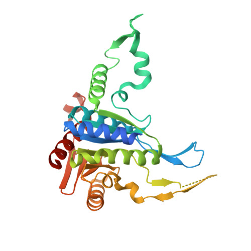
    The structure of human phosphopantothenoylcysteine (PPC) synthetase was determined at 2.3 A resolution. PPC synthetase is a dimer with identical monomers. Some features of the monomer fold resemble a group of NAD-dependent enzymes, while other features resemble the ribokinase fold. The ATP, phosphopantothenate, and cysteine binding sites were deduced from modeling studies. Highly conserved ATP binding residues include Gly43, Ser61, Gly63, Gly66, Phe230, and Asn258. Highly conserved phosphopantothenate binding residues include Asn59, Ala179, Ala180, and Asp183 from one monomer and Arg55' from the adjacent monomer. The structure predicts a ping pong mechanism with initial formation of an acyladenylate intermediate, followed by release of pyrophosphate and attack by cysteine to form the final products PPC and AMP.


  • Organizational Affiliation

    Department of Chemistry and Chemical Biology, Cornell University, Ithaca, NY 14853, USA.


Macromolecules
Find similar proteins by:  (by identity cutoff)  |  3D Structure
Entity ID: 1
MoleculeChains Sequence LengthOrganismDetailsImage
Phosphopantothenoylcysteine synthetase
A, B
313Homo sapiensMutation(s): 0 
Gene Names: coaB
EC: 6.3.2.5
UniProt & NIH Common Fund Data Resources
Find proteins for Q9HAB8 (Homo sapiens)
Explore Q9HAB8 
Go to UniProtKB:  Q9HAB8
PHAROS:  Q9HAB8
GTEx:  ENSG00000127125 
Entity Groups  
Sequence Clusters30% Identity50% Identity70% Identity90% Identity95% Identity100% Identity
UniProt GroupQ9HAB8
Sequence Annotations
Expand
  • Reference Sequence
Experimental Data & Validation

Experimental Data

  • Method: X-RAY DIFFRACTION
  • Resolution: 2.30 Å
  • R-Value Free: 0.272 
  • R-Value Work: 0.229 
  • R-Value Observed: 0.229 
  • Space Group: P 43 21 2
Unit Cell:
Length ( Å )Angle ( ˚ )
a = 73.16α = 90
b = 73.16β = 90
c = 281.83γ = 90
Software Package:
Software NamePurpose
CNSrefinement
DENZOdata reduction
CCP4data scaling
CNSphasing

Structure Validation

View Full Validation Report


Entry History 

Deposition Data

Revision History  (Full details and data files)

  • Version 1.0: 2003-09-02
    Type: Initial release
  • Version 1.1: 2008-04-29
    Changes: Version format compliance
  • Version 1.2: 2011-07-13
    Changes: Version format compliance
  • Version 1.3: 2024-02-14
    Changes: Data collection, Database references, Derived calculations
  • Version 1.4: 2024-04-03
    Changes: Refinement description