1OLA

THE STRUCTURAL BASIS OF MULTISPECIFICITY IN THE OLIGOPEPTIDE-BINDING PROTEIN OPPA


Experimental Data Snapshot

  • Method: X-RAY DIFFRACTION
  • Resolution: 2.10 Å
  • R-Value Observed: 0.167 

wwPDB Validation   3D Report Full Report


This is version 1.2 of the entry. See complete history


Literature

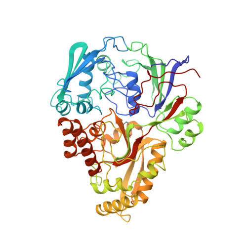
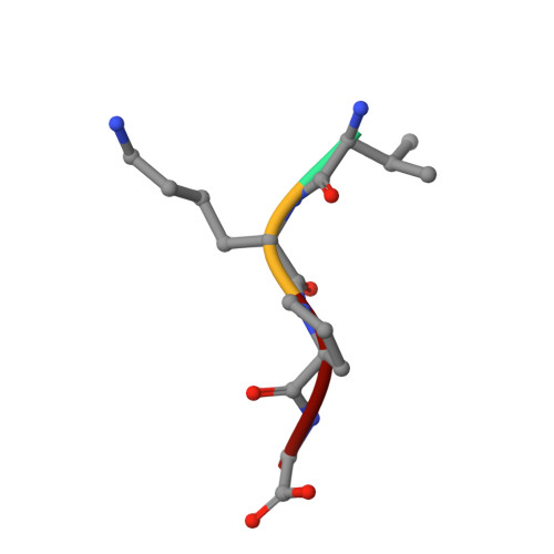
The structural basis of sequence-independent peptide binding by OppA protein.

Tame, J.R.Murshudov, G.N.Dodson, E.J.Neil, T.K.Dodson, G.G.Higgins, C.F.Wilkinson, A.J.

(1994) Science 264: 1578-1581

  • DOI: https://doi.org/10.1126/science.8202710
  • Primary Citation of Related Structures:  
    1OLA

  • PubMed Abstract: 

    Specific protein-ligand interactions are critical for cellular function, and most proteins select their partners with sharp discrimination. However, the oligopeptide-binding protein of Salmonella typhimurium (OppA) binds peptides of two to five amino acid residues without regard to sequence. The crystal structure of OppA reveals a three-domain organization, unlike other periplasmic binding proteins. In OppA-peptide complexes, the ligands are completely enclosed in the protein interior, a mode of binding that normally imposes tight specificity. The protein fulfills the hydrogen bonding and electrostatic potential of the ligand main chain and accommodates the peptide side chains in voluminous hydrated cavities.


  • Organizational Affiliation

    Department of Chemistry, University of York, UK.


Macromolecules
Find similar proteins by:  (by identity cutoff)  |  3D Structure
Entity ID: 1
MoleculeChains Sequence LengthOrganismDetailsImage
OLIGO-PEPTIDE BINDING PROTEIN517Salmonella enterica subsp. enterica serovar TyphimuriumMutation(s): 0 
UniProt
Find proteins for P06202 (Salmonella typhimurium (strain LT2 / SGSC1412 / ATCC 700720))
Explore P06202 
Go to UniProtKB:  P06202
Entity Groups  
Sequence Clusters30% Identity50% Identity70% Identity90% Identity95% Identity100% Identity
UniProt GroupP06202
Sequence Annotations
Expand
  • Reference Sequence

Find similar proteins by:  Sequence   |   3D Structure  

Entity ID: 2
MoleculeChains Sequence LengthOrganismDetailsImage
PEPTIDE VAL-LYS-PRO-GLY4N/AMutation(s): 0 
Sequence Annotations
Expand
  • Reference Sequence
Small Molecules
Ligands 1 Unique
IDChains Name / Formula / InChI Key2D Diagram3D Interactions
IUM
Query on IUM

Download Ideal Coordinates CCD File 
C [auth A],
D [auth A]
URANYL (VI) ION
O2 U
IZLHQJIUOZHTLW-UHFFFAOYSA-N
Experimental Data & Validation

Experimental Data

  • Method: X-RAY DIFFRACTION
  • Resolution: 2.10 Å
  • R-Value Observed: 0.167 
  • Space Group: P 21 21 2
Unit Cell:
Length ( Å )Angle ( ˚ )
a = 106.5α = 90
b = 74.5β = 90
c = 70γ = 90
Software Package:
Software NamePurpose
PROLSQrefinement

Structure Validation

View Full Validation Report


Entry History 

Deposition Data

Revision History  (Full details and data files)

  • Version 1.0: 1994-07-31
    Type: Initial release
  • Version 1.1: 2008-03-24
    Changes: Version format compliance
  • Version 1.2: 2011-07-13
    Changes: Version format compliance