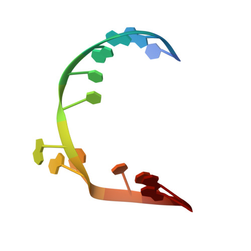
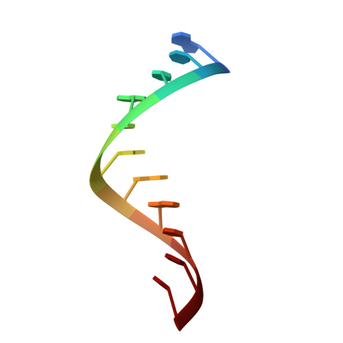
Crystal structure of the leadzyme at 1.8 A resolution: metal ion binding and the implications for catalytic mechanism and allo site ion regulation.
Wedekind, J.E., McKay, D.B.(2003) Biochemistry 42: 9554-9563
- PubMed: 12911297
- DOI: https://doi.org/10.1021/bi0300783
- Primary Citation of Related Structures:
1NUJ, 1NUV - PubMed Abstract:
The leadzyme is a small ribozyme, derived from in vitro selection, which catalyzes site specific, Pb(2+)-dependent RNA cleavage. Pb(2+) is required for activity; Mg(2+) inhibits activity, while many divalent and trivalent ions enhance it. The leadzyme structure consists of an RNA duplex interrupted by a trinucleotide bulge. Here, crystal structures determined to 1.8 A resolution, both with Mg(2+) as the sole divalent counterion and with Mg(2+) and Sr(2+) (which mimics Pb(2+) with respect to binding but not catalysis), reveal the metal ion interactions with both the ground state and precatalytic conformations of the leadzyme. Mg(H(2)O)(6)(2+) ions bridge complementary strands of the duplex at multiple locations by binding tandem purines of one RNA strand in the major groove. At one site, Mg(H(2)O)(6)(2+) ligates the phosphodiester backbone of the trinucleotide bulge in the ground state conformation, but not in the precatalytic conformation, suggesting (a) Mg(2+) may inhibit leadzyme activity by stabilizing the ground state and (b) metal ions which displace Mg(2+) from this site may activate the leadzyme. Binding of Sr(2+) to the presumed catalytic Pb(2+) site in the precatalytic leadzyme induces local structural changes in a manner that would facilitate alignment of the catalytic ribose 2'-hydroxyl with the scissile bond for cleavage. These data support a model wherein binding of a catalytic ion to a precatalytic conformation of the leadzyme, in conjunction with the flexibility of the trinucleotide bulge, may facilitate structural rearrangements around the scissle phosphodiester bond favoring configurations that allow bond cleavage.
Organizational Affiliation:
Department of Structural Biology, Stanford University School of Medicine, Stanford, California 94305, USA.