Heme-assisted S-nitrosation of a proximal thiolate in a nitric oxide transport protein.
Weichsel, A., Maes, E.M., Andersen, J.F., Valenzuela, J.G., Shokhireva, T.K., Walker, F.A., Montfort, W.R.(2005) Proc Natl Acad Sci U S A 102: 594-599
- PubMed: 15637157
- DOI: https://doi.org/10.1073/pnas.0406549102
- Primary Citation of Related Structures:
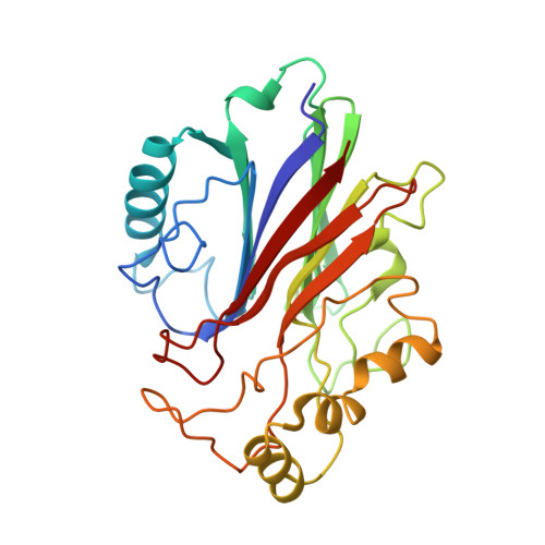
1NTF, 1Y21 - PubMed Abstract:
Certain bloodsucking insects deliver nitric oxide (NO) while feeding, to induce vasodilation and inhibit blood coagulation. We have expressed, characterized, and determined the crystal structure of the Cimex lectularius (bedbug) nitrophorin, the protein responsible for NO storage and delivery, to understand how the insect successfully handles this reactive molecule. Surprisingly, NO binds not only to the ferric nitrophorin heme, but it can also be stored as an S-nitroso (SNO) conjugate of the proximal heme cysteine (Cys-60) when present at higher concentrations. EPR- and UV-visible spectroscopies, and a crystallographic structure determination to 1.75-A resolution, reveal SNO formation to proceed with reduction of the heme iron, yielding an Fe-NO complex. Stopped-flow kinetic measurements indicate that an ordered reaction mechanism takes place: initial NO binding occurs at the ferric heme and is followed by heme reduction, Cys-60 release from the heme iron, and SNO formation. Release of NO occurs through a reversal of these steps. These data provide, to our knowledge, the first view of reversible metal-assisted SNO formation in a protein and suggest a mechanism for its role in NO release from ferrous heme. This mechanism and Cimex nitrophorin structure are completely unlike those of the nitrophorins from Rhodnius prolixus, where NO protection is provided by a large conformational change that buries the heme nitrosyl complex, highlighting the remarkable evolution of proteins that assist insects in bloodfeeding.
Organizational Affiliation:
Department of Biochemistry and Molecular Biophysics, University of Arizona, Tucson, AZ 85721, USA.