1M6I

Crystal Structure of Apoptosis Inducing Factor (AIF)


Experimental Data Snapshot

  • Method: X-RAY DIFFRACTION
  • Resolution: 1.80 Å
  • R-Value Free: 0.241 
  • R-Value Work: 0.225 

wwPDB Validation   3D Report Full Report


Ligand Structure Quality Assessment 


This is version 1.3 of the entry. See complete history

Re-refinement Note

A newer entry is available that reflects an alternative modeling of the original data: 4BV6


Literature

DNA binding is required for the apoptogenic action of apoptosis inducing factor.

Ye, H.Cande, C.Stephanou, N.C.Jiang, S.Gurbuxani, S.Larochette, N.Daugas, E.Garrido, C.Kroemer, G.Wu, H.

(2002) Nat Struct Biol 9: 680-684

  • DOI: https://doi.org/10.1038/nsb836
  • Primary Citation of Related Structures:  
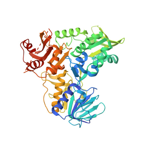
    1M6I

  • PubMed Abstract: 

    The execution of apoptosis or programmed cell death comprises both caspase-dependent and caspase-independent processes. Apoptosis inducing factor (AIF) was identified as a major player in caspase-independent cell death. It induces chromatin condensation and initial DNA cleavage via an unknown molecular mechanism. Here we report the crystal structure of human AIF at 1.8 A resolution. The structure reveals the presence of a strong positive electrostatic potential at the AIF surface, although the calculated isoelectric point for the entire protein is neutral. We show that recombinant AIF interacts with DNA in a sequence-independent manner. In addition, in cells treated with an apoptotic stimulus, endogenous AIF becomes co-localized with DNA at an early stage of nuclear morphological changes. Structure-based mutagenesis shows that DNA-binding defective mutants of AIF fail to induce cell death while retaining nuclear translocation. The potential DNA-binding site identified from mutagenesis also coincides with computational docking of a DNA duplex. These observations suggest that AIF-induced nuclear apoptosis requires a direct interaction with DNA.


  • Organizational Affiliation

    Department of Biochemistry, Weill Medical College of Cornell University, 1300 York Avenue, New York, New York 10021, USA.


Macromolecules
Find similar proteins by:  (by identity cutoff)  |  3D Structure
Entity ID: 1
MoleculeChains Sequence LengthOrganismDetailsImage
Programmed cell death protein 8493Homo sapiensMutation(s): 0 
UniProt & NIH Common Fund Data Resources
Find proteins for O95831 (Homo sapiens)
Explore O95831 
Go to UniProtKB:  O95831
PHAROS:  O95831
GTEx:  ENSG00000156709 
Entity Groups  
Sequence Clusters30% Identity50% Identity70% Identity90% Identity95% Identity100% Identity
UniProt GroupO95831
Sequence Annotations
Expand
  • Reference Sequence
Small Molecules
Ligands 1 Unique
IDChains Name / Formula / InChI Key2D Diagram3D Interactions
FAD
Query on FAD

Download Ideal Coordinates CCD File 
B [auth A]FLAVIN-ADENINE DINUCLEOTIDE
C27 H33 N9 O15 P2
VWWQXMAJTJZDQX-UYBVJOGSSA-N
Experimental Data & Validation

Experimental Data

  • Method: X-RAY DIFFRACTION
  • Resolution: 1.80 Å
  • R-Value Free: 0.241 
  • R-Value Work: 0.225 
  • Space Group: P 1 21 1
Unit Cell:
Length ( Å )Angle ( ˚ )
a = 50.9α = 90
b = 90.5β = 94.6
c = 60.5γ = 90
Software Package:
Software NamePurpose
HKL-2000data collection
SCALEPACKdata scaling
SOLVEphasing
CNSrefinement
HKL-2000data reduction

Structure Validation

View Full Validation Report


Ligand Structure Quality Assessment 


Entry History 

Deposition Data

Revision History  (Full details and data files)

  • Version 1.0: 2002-08-28
    Type: Initial release
  • Version 1.1: 2008-04-28
    Changes: Version format compliance
  • Version 1.2: 2011-07-13
    Changes: Version format compliance
  • Version 1.3: 2024-02-14
    Changes: Data collection, Database references, Derived calculations