1KCK

Bacillus circulans strain 251 Cyclodextrin glycosyl transferase mutant N193G


Experimental Data Snapshot

  • Method: X-RAY DIFFRACTION
  • Resolution: 2.43 Å
  • R-Value Free: 0.205 
  • R-Value Work: 0.159 

wwPDB Validation   3D Report Full Report


Ligand Structure Quality Assessment 


This is version 3.2 of the entry. See complete history


Literature

The remote substrate binding subsite -6 in cyclodextrin-glycosyltransferase controls the transferase activity of the enzyme via an induced-fit mechanism.

Leemhuis, H.Uitdehaag, J.C.Rozeboom, H.J.Dijkstra, B.W.Dijkhuizen, L.

(2002) J Biol Chem 277: 1113-1119

  • DOI: https://doi.org/10.1074/jbc.M106667200
  • Primary Citation of Related Structures:  
    1KCK, 1KCL

  • PubMed Abstract: 

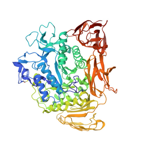
    Cyclodextrin-glycosyltransferase (CGTase) catalyzes the formation of alpha-, beta-, and gamma-cyclodextrins (cyclic alpha-(1,4)-linked oligosaccharides of 6, 7, or 8 glucose residues, respectively) from starch. Nine substrate binding subsites were observed in an x-ray structure of the CGTase from Bacillus circulans strain 251 complexed with a maltononaose substrate. Subsite -6 is conserved in CGTases, suggesting its importance for the reactions catalyzed by the enzyme. To investigate this in detail, we made six mutant CGTases (Y167F, G179L, G180L, N193G, N193L, and G179L/G180L). All subsite -6 mutants had decreased k(cat) values for beta-cyclodextrin formation, as well as for the disproportionation and coupling reactions, but not for hydrolysis. Especially G179L, G180L, and G179L/G180L affected the transglycosylation activities, most prominently for the coupling reactions. The results demonstrate that (i) subsite -6 is important for all three CGTase-catalyzed transglycosylation reactions, (ii) Gly-180 is conserved because of its importance for the circularization of the linear substrates, (iii) it is possible to independently change cyclization and coupling activities, and (iv) substrate interactions at subsite -6 activate the enzyme in catalysis via an induced-fit mechanism. This article provides for the first time definite biochemical evidence for such an induced-fit mechanism in the alpha-amylase family.


  • Organizational Affiliation

    Department of Microbiology, Groningen Biomolecular Sciences and Biotechnology Institute, University of Groningen, Kerklaan 30, 9751 NN Haren, The Netherlands.


Macromolecules
Find similar proteins by:  (by identity cutoff)  |  3D Structure
Entity ID: 1
MoleculeChains Sequence LengthOrganismDetailsImage
CYCLODEXTRIN GLYCOSYLTRANSFERASE686Niallia circulansMutation(s): 1 
EC: 2.4.1.19
UniProt
Find proteins for P43379 (Niallia circulans)
Explore P43379 
Go to UniProtKB:  P43379
Entity Groups  
Sequence Clusters30% Identity50% Identity70% Identity90% Identity95% Identity100% Identity
UniProt GroupP43379
Sequence Annotations
Expand
  • Reference Sequence
Oligosaccharides

Help

Entity ID: 2
MoleculeChains Length2D Diagram Glycosylation3D Interactions
alpha-D-quinovopyranose-(1-4)-alpha-D-glucopyranose
B
2N/A
Glycosylation Resources
GlyTouCan:  G96906VH
GlyCosmos:  G96906VH
Entity ID: 3
MoleculeChains Length2D Diagram Glycosylation3D Interactions
alpha-D-glucopyranose-(1-4)-alpha-D-glucopyranose
C
2N/A
Glycosylation Resources
GlyTouCan:  G07411ON
GlyCosmos:  G07411ON
Entity ID: 4
MoleculeChains Length2D Diagram Glycosylation3D Interactions
alpha-D-glucopyranose-(1-4)-alpha-D-glucopyranose-(1-4)-alpha-D-glucopyranose
D
3N/A
Glycosylation Resources
GlyTouCan:  G96370VA
GlyCosmos:  G96370VA
GlyGen:  G96370VA
Entity ID: 5
MoleculeChains Length2D Diagram Glycosylation3D Interactions
alpha-D-glucopyranose-(1-4)-alpha-D-glucopyranose-(1-4)-beta-D-glucopyranose
E
3N/A
Glycosylation Resources
GlyTouCan:  G00395FS
GlyCosmos:  G00395FS
GlyGen:  G00395FS
Experimental Data & Validation

Experimental Data

  • Method: X-RAY DIFFRACTION
  • Resolution: 2.43 Å
  • R-Value Free: 0.205 
  • R-Value Work: 0.159 
  • Space Group: P 21 21 21
Unit Cell:
Length ( Å )Angle ( ˚ )
a = 120.686α = 90
b = 111.162β = 90
c = 65.944γ = 90
Software Package:
Software NamePurpose
XDSdata scaling
XDSdata reduction
CNSrefinement
CNSphasing

Structure Validation

View Full Validation Report


Ligand Structure Quality Assessment 


Entry History 

Deposition Data

Revision History  (Full details and data files)

  • Version 1.0: 2002-01-16
    Type: Initial release
  • Version 1.1: 2008-04-27
    Changes: Version format compliance
  • Version 1.2: 2011-07-13
    Changes: Version format compliance
  • Version 2.0: 2017-10-25
    Changes: Atomic model, Derived calculations, Structure summary
  • Version 3.0: 2020-07-29
    Type: Remediation
    Reason: Carbohydrate remediation
    Changes: Atomic model, Data collection, Derived calculations, Structure summary
  • Version 3.1: 2021-10-27
    Changes: Database references, Structure summary
  • Version 3.2: 2023-08-16
    Changes: Data collection, Refinement description