1KC6

HincII Bound to Cognate DNA


Experimental Data Snapshot

  • Method: X-RAY DIFFRACTION
  • Resolution: 2.60 Å
  • R-Value Free: 0.285 
  • R-Value Work: 0.219 

wwPDB Validation   3D Report Full Report


This is version 1.3 of the entry. See complete history


Literature

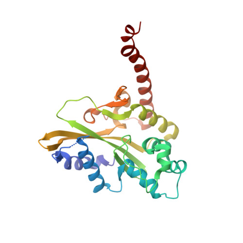
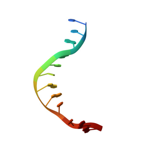
Sequence selectivity and degeneracy of a restriction endonuclease mediated by DNA intercalation.

Horton, N.C.Dorner, L.F.Perona, J.J.

(2002) Nat Struct Biol 9: 42-47

  • DOI: https://doi.org/10.1038/nsb741
  • Primary Citation of Related Structures:  
    1KC6

  • PubMed Abstract: 

    The crystal structure of the HincII restriction endonuclease-DNA complex shows that degenerate specificity for blunt-ended cleavage at GTPyPuAC sequences arises from indirect readout of conformational preferences at the center pyrimidine-purine step. Protein-induced distortion of the DNA is accomplished by intercalation of glutamine side chains into the major groove on either side of the recognition site, generating bending by either tilt or roll at three distinct loci. The intercalated side chains propagate a concerted shift of all six target-site base pairs toward the minor groove, producing an unusual cross-strand purine stacking at the center pyrimidine-purine step. Comparison of the HincII and EcoRV cocrystal structures suggests that sequence-dependent differences in base-stacking free energies are a crucial underlying factor mediating protein recognition by indirect readout.


  • Organizational Affiliation

    Department of Chemistry and Biochemistry, and Interdepartmental Program in Biomolecular Science and Engineering, University of California at Santa Barbara, Santa Barbara, California 93106-9510, USA.


Macromolecules

Find similar proteins by:  (by identity cutoff)  |  3D Structure
Entity ID: 2
MoleculeChains Sequence LengthOrganismDetailsImage
TYPE II RESTRICTION ENZYME HINCIIE [auth A],
F [auth B],
G [auth C],
H [auth D]
257Haemophilus influenzaeMutation(s): 0 
EC: 3.1.21.4
UniProt
Find proteins for P17743 (Haemophilus influenzae)
Explore P17743 
Go to UniProtKB:  P17743
Entity Groups  
Sequence Clusters30% Identity50% Identity70% Identity90% Identity95% Identity100% Identity
UniProt GroupP17743
Sequence Annotations
Expand
  • Reference Sequence

Find similar nucleic acids by:  Sequence   |   3D Structure  

Entity ID: 1
MoleculeChains LengthOrganismImage
5'-D(P*CP*CP*GP*GP*TP*CP*GP*AP*CP*CP*GP*G)-3'A [auth E],
B [auth F],
C [auth G],
D [auth H]
12N/A
Sequence Annotations
Expand
  • Reference Sequence
Small Molecules
Ligands 1 Unique
IDChains Name / Formula / InChI Key2D Diagram3D Interactions
NA
Query on NA

Download Ideal Coordinates CCD File 
I [auth E],
J [auth A]
SODIUM ION
Na
FKNQFGJONOIPTF-UHFFFAOYSA-N
Experimental Data & Validation

Experimental Data

  • Method: X-RAY DIFFRACTION
  • Resolution: 2.60 Å
  • R-Value Free: 0.285 
  • R-Value Work: 0.219 
  • Space Group: I 21 21 21
Unit Cell:
Length ( Å )Angle ( ˚ )
a = 67.12α = 90
b = 177.67β = 90
c = 256.36γ = 90
Software Package:
Software NamePurpose
SOLVEphasing
CNSrefinement
MOSFLMdata reduction
CCP4data scaling

Structure Validation

View Full Validation Report


Entry History 

Deposition Data

Revision History  (Full details and data files)

  • Version 1.0: 2001-12-07
    Type: Initial release
  • Version 1.1: 2008-05-05
    Changes: Version format compliance
  • Version 1.2: 2011-07-13
    Changes: Version format compliance
  • Version 1.3: 2024-02-07
    Changes: Data collection, Database references, Derived calculations