1IY1

Crystal structure of the FtsH ATPase domain with ADP from Thermus thermophilus


Experimental Data Snapshot

  • Method: X-RAY DIFFRACTION
  • Resolution: 2.80 Å
  • R-Value Free: 0.278 
  • R-Value Work: 0.207 

wwPDB Validation   3D Report Full Report


Ligand Structure Quality Assessment 


This is version 1.3 of the entry. See complete history


Literature

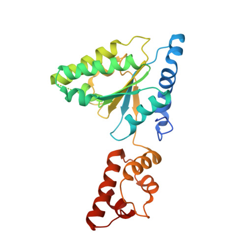
Hexameric ring structure of the ATPase domain of the membrane-integrated metalloprotease FtsH from Thermus thermophilus HB8

Niwa, H.Tsuchiya, D.Makyio, H.Yoshida, M.Morikawa, K.

(2002) Structure 10: 1415-1423

  • DOI: https://doi.org/10.1016/s0969-2126(02)00855-9
  • Primary Citation of Related Structures:  
    1IXZ, 1IY0, 1IY1, 1IY2

  • PubMed Abstract: 

    FtsH is a cytoplasmic membrane-integrated, ATP-dependent metalloprotease, which processively degrades both cytoplasmic and membrane proteins in concert with unfolding. The FtsH protein is divided into the N-terminal transmembrane region and the larger C-terminal cytoplasmic region, which consists of an ATPase domain and a protease domain. We have determined the crystal structures of the Thermus thermophilus FtsH ATPase domain in the nucleotide-free and AMP-PNP- and ADP-bound states, in addition to the domain with the extra preceding segment. Combined with the mapping of the putative substrate binding region, these structures suggest that FtsH internally forms a hexameric ring structure, in which ATP binding could cause a conformational change to facilitate transport of substrates into the protease domain through the central pore.


  • Organizational Affiliation

    Department of Structural Biology, Biomolecular Engineering Research Institute, 6-2-3 Furuedai, Suita, Osaka, Japan.


Macromolecules
Find similar proteins by:  (by identity cutoff)  |  3D Structure
Entity ID: 1
MoleculeChains Sequence LengthOrganismDetailsImage
ATP-dependent metalloprotease FtsH254Thermus thermophilusMutation(s): 0 
Gene Names: FtsH
UniProt
Find proteins for Q5SI82 (Thermus thermophilus (strain ATCC 27634 / DSM 579 / HB8))
Explore Q5SI82 
Go to UniProtKB:  Q5SI82
Entity Groups  
Sequence Clusters30% Identity50% Identity70% Identity90% Identity95% Identity100% Identity
UniProt GroupQ5SI82
Sequence Annotations
Expand
  • Reference Sequence
Small Molecules
Ligands 1 Unique
IDChains Name / Formula / InChI Key2D Diagram3D Interactions
ADP
Query on ADP

Download Ideal Coordinates CCD File 
B [auth A]ADENOSINE-5'-DIPHOSPHATE
C10 H15 N5 O10 P2
XTWYTFMLZFPYCI-KQYNXXCUSA-N
Experimental Data & Validation

Experimental Data

  • Method: X-RAY DIFFRACTION
  • Resolution: 2.80 Å
  • R-Value Free: 0.278 
  • R-Value Work: 0.207 
  • Space Group: P 65
Unit Cell:
Length ( Å )Angle ( ˚ )
a = 73.875α = 90
b = 73.875β = 90
c = 84.611γ = 120
Software Package:
Software NamePurpose
DENZOdata reduction
SCALEPACKdata scaling
AMoREphasing
CNSrefinement

Structure Validation

View Full Validation Report


Ligand Structure Quality Assessment 


Entry History 

Deposition Data

Revision History  (Full details and data files)

  • Version 1.0: 2002-11-06
    Type: Initial release
  • Version 1.1: 2008-04-27
    Changes: Version format compliance
  • Version 1.2: 2011-07-13
    Changes: Version format compliance
  • Version 1.3: 2023-12-27
    Changes: Data collection, Database references, Derived calculations