Crystal structure and dimerization equilibria of PcoC, a methionine-rich copper resistance protein from Escherichia coli
Wernimont, A.K., Huffman, D.L., Finney, L.A., Demeler, B., O'Halloran, T.V., Rosenzweig, A.C.(2003) J Biol Inorg Chem 8: 185-194
- PubMed: 12459914
- DOI: https://doi.org/10.1007/s00775-002-0404-9
- Primary Citation of Related Structures:
1IX2, 1LYQ - PubMed Abstract:
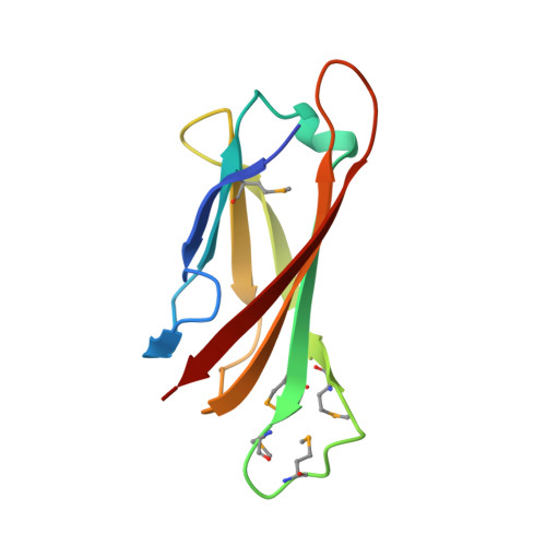
PcoC is a soluble periplasmic protein encoded by the plasmid-born pco copper resistance operon of Escherichia coli. Like PcoA, a multicopper oxidase encoded in the same locus and its chromosomal homolog CueO, PcoC contains unusual methionine rich sequences. Although essential for copper resistance, the functions of PcoC, PcoA, and their conserved methionine-rich sequences are not known. Similar methionine motifs observed in eukaryotic copper transporters have been proposed to bind copper, but there are no precedents for such metal binding sites in structurally characterized proteins. The high-resolution structures of apo PcoC, determined for both the native and selenomethionine-containing proteins, reveal a seven-stranded beta barrel with the methionines unexpectedly housed on a solvent-exposed loop. Several potential metal-binding sites can be discerned by comparing the structures to spectroscopic data reported for copper-loaded PcoC. In the native structure, the methionine loop interacts with the same loop on a second molecule in the asymmetric unit. In the selenomethionine structure, the methionine loops are more exposed, forming hydrophobic patches on the protein surface. These two arrangements suggest that the methionine motifs might function in protein-protein interactions between PcoC molecules or with other methionine-rich proteins such as PcoA. Analytical ultracentrifugation data indicate that a weak monomer-dimer equilibrium exists in solution for the apo protein. Dimerization is significantly enhanced upon binding Cu(I) with a measured delta(deltaG degrees )
Organizational Affiliation:
Department of Biochemistry, Northwestern University, Evanston, IL 60208, USA.