1IB0

CRYSTAL STRUCTURE OF RAT B5R IN COMPLEX WITH FAD AND NAD


Experimental Data Snapshot

  • Method: X-RAY DIFFRACTION
  • Resolution: 2.30 Å
  • R-Value Free: 0.244 
  • R-Value Work: 0.218 

wwPDB Validation   3D Report Full Report


This is version 1.3 of the entry. See complete history


Literature

The structure and biochemistry of NADH-dependent cytochrome b5 reductase are now consistent.

Bewley, M.C.Marohnic, C.C.Barber, M.J.

(2001) Biochemistry 40: 13574-13582

  • DOI: https://doi.org/10.1021/bi0106336
  • Primary Citation of Related Structures:  
    1I7P, 1IB0

  • PubMed Abstract: 

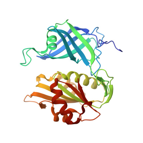
    Cytochrome b5 reductase (cb5r) (EC 1.6.6.2) catalyzes the reduction of two molecules of cytochrome b5 using NADH as the physiological electron donor. The structure of pig cb5r at 2.4 A resolution was previously reported in the literature, but it was inconsistent with the biochemistry; for example, K83 and C245 were both implicated in the mechanism, but were not located at the active site. To address this problem, we have determined the structures of cb5r from rat at 2.0 A resolution and in a complex with NAD+ at 2.3 A resolution. We found significant differences throughout the rat structure compared to that of pig, including the locations of the lysine and cysteine residues mentioned above. To test the structural models, we made single amino acid substitutions of this lysine and showed that all substitutions produced correctly folded proteins and exhibited normal flavin behavior. However, the apparent kcat(NADH) decreased, and the apparent K(m) for NADH increased; the K(m)'s for cytochrome b5 were unchanged relative to that of the wild type. The largest effect was for the glutamate-substituted protein, which was further characterized using a charge transfer assay and found to be less efficient at NADH utilization than the wild type. These results are consistent with a role for this lysine in stabilizing the NADH-bound form of cb5r. We have concluded that the pig structure was mistraced in several regions and have reinterpreted mutants in these regions that give rise to the hereditary disease methemoglobinemia.


  • Organizational Affiliation

    Biology Department, Brookhaven National Laboratory, Upton, New York 11973, USA. bewley@bnl.gov


Macromolecules
Find similar proteins by:  (by identity cutoff)  |  3D Structure
Entity ID: 1
MoleculeChains Sequence LengthOrganismDetailsImage
NADH-CYTOCHROME B5 REDUCTASE274Rattus norvegicusMutation(s): 0 
Gene Names: DIA1
EC: 1.6.2.2
UniProt
Find proteins for P20070 (Rattus norvegicus)
Explore P20070 
Go to UniProtKB:  P20070
Entity Groups  
Sequence Clusters30% Identity50% Identity70% Identity90% Identity95% Identity100% Identity
UniProt GroupP20070
Sequence Annotations
Expand
  • Reference Sequence
Small Molecules
Ligands 2 Unique
IDChains Name / Formula / InChI Key2D Diagram3D Interactions
FAD
Query on FAD

Download Ideal Coordinates CCD File 
B [auth A]FLAVIN-ADENINE DINUCLEOTIDE
C27 H33 N9 O15 P2
VWWQXMAJTJZDQX-UYBVJOGSSA-N
NAD
Query on NAD

Download Ideal Coordinates CCD File 
C [auth A]NICOTINAMIDE-ADENINE-DINUCLEOTIDE
C21 H27 N7 O14 P2
BAWFJGJZGIEFAR-NNYOXOHSSA-N
Experimental Data & Validation

Experimental Data

  • Method: X-RAY DIFFRACTION
  • Resolution: 2.30 Å
  • R-Value Free: 0.244 
  • R-Value Work: 0.218 
  • Space Group: P 21 21 21
Unit Cell:
Length ( Å )Angle ( ˚ )
a = 67.612α = 90
b = 69.458β = 90
c = 78.787γ = 90
Software Package:
Software NamePurpose
AMoREphasing
CNSrefinement
DENZOdata reduction
SCALEPACKdata scaling

Structure Validation

View Full Validation Report


Entry History 

Deposition Data

Revision History  (Full details and data files)

  • Version 1.0: 2001-12-12
    Type: Initial release
  • Version 1.1: 2008-04-27
    Changes: Version format compliance
  • Version 1.2: 2011-07-13
    Changes: Version format compliance
  • Version 1.3: 2023-08-09
    Changes: Data collection, Database references, Derived calculations, Refinement description