Tyrosine hydrogen bonds make a large contribution to protein stability.
Pace, C.N., Horn, G., Hebert, E.J., Bechert, J., Shaw, K., Urbanikova, L., Scholtz, J.M., Sevcik, J.(2001) J Mol Biol 312: 393-404
- PubMed: 11554795
- DOI: https://doi.org/10.1006/jmbi.2001.4956
- Primary Citation of Related Structures:
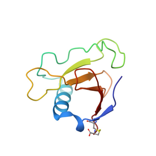
1I70, 1I8V - PubMed Abstract:
The aim of this study was to gain a better understanding of the contribution of hydrogen bonds by tyrosine -OH groups to protein stability. The amino acid sequences of RNases Sa and Sa3 are 69 % identical and each contains eight Tyr residues with seven at equivalent structural positions. We have measured the stability of the 16 tyrosine to phenylalanine mutants. For two equivalent mutants, the stability increases by 0.3 kcal/mol (RNase Sa Y30F) and 0.5 kcal/mol (RNase Sa3 Y33F) (1 kcal=4.184 kJ). For all of the other mutants, the stability decreases with the greatest decrease being 3.6 kcal/mol for RNase Sa Y52F. Seven of the 16 tyrosine residues form intramolecular hydrogen bonds and the average decrease in stability for these is 2.0(+/-1.0) kcal/mol. For the nine tyrosine residues that do not form intramolecular hydrogen bonds, the average decrease in stability is 0.4(+/-0.6) kcal/mol. Thus, most tyrosine -OH groups contribute favorably to protein stability even if they do not form intramolecular hydrogen bonds. Generally, the stability changes for equivalent positions in the two proteins are remarkably similar. Crystal structures were determined for two of the tyrosine to phenylalanine mutants of RNase Sa: Y80F (1.2 A), and Y86F (1.7 A). The structures are very similar to that of wild-type RNase Sa, and the hydrogen bonding partners of the tyrosine residues always form intermolecular hydrogen bonds to water in the mutants. These results provide further evidence that the hydrogen bonding and van der Waals interactions of polar groups in the tightly packed interior of folded proteins are more favorable than similar interactions with water in the unfolded protein, and that polar group burial makes a substantial contribution to protein stability.
Organizational Affiliation:
Department of Medical Biochemistry and Genetics, Texas A&M University, College Station, 77843-1114, USA. nickpace@tamu.edu