1H9L

PORCINE PANCREATIC ELASTASE COMPLEXED WITH ACETYL-VAL-GLU-PRO-ILE-COOH


Experimental Data Snapshot

  • Method: X-RAY DIFFRACTION
  • Resolution: 1.67 Å
  • R-Value Free: 0.199 
  • R-Value Work: 0.158 
  • R-Value Observed: 0.159 

wwPDB Validation   3D Report Full Report


This is version 1.3 of the entry. See complete history


Literature

Kinetic and Crystallographic Analysis of Complexes Formed between Elastase and Peptides from Beta-Casein

Wright, P.A.Wilmouth, R.C.Clifton, I.J.Schofield, C.J.

(2001) Eur J Biochem 268: 2969

  • DOI: https://doi.org/10.1046/j.1432-1327.2001.02186.x
  • Primary Citation of Related Structures:  
    1H9L

  • PubMed Abstract: 

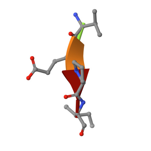
    Human beta-casomorphin-7 (NH2-Tyr-Pro-Phe-Val-Glu-Pro-Ile-CO2H) is a naturally occurring peptide inhibitor of elastase that has been shown to form an acyl-enzyme complex stable enough for X-ray crystallographic analysis at pH 5. To investigate the importance of the N-terminal residues of the beta-casomorphin-7 peptide for the inhibition of elastase, kinetic and crystallographic analyses were undertaken to identify the minimum number of residues required for effective formation of a stable complex between truncated beta-casomorphin-7 peptides and porcine pancreatic elastase (PPE). The results clearly demonstrate that significant inhibition of PPE can be effected by simple tri-, tetra-and pentapeptides terminating in a carboxylic acid. These results also suggest that in vivo regulation of protease activity could be mediated via short peptides as well as by proteins. Crystallographic analysis of the complex formed between N-acetyl-Val-Glu-Pro-Ile-CO2H and PPE at pH 5 (to 1.67 A resolution) revealed an active site water molecule in an analogous position to that observed in the PPE/beta-casomorphin-7 structure supportive of its assignment as the 'hydrolytic water' in the deacylation step of serine protease catalysis.


  • Organizational Affiliation

    The Dyson Perrins Laboratory and The Oxford Centre for Molecular Sciences, Oxford, UK.


Macromolecules

Find similar proteins by:  Sequence   |   3D Structure  

Entity ID: 1
MoleculeChains Sequence LengthOrganismDetailsImage
PEPTIDE INHIBITOR5synthetic constructMutation(s): 0 
Sequence Annotations
Expand
  • Reference Sequence
Find similar proteins by:  (by identity cutoff)  |  3D Structure
Entity ID: 2
MoleculeChains Sequence LengthOrganismDetailsImage
ELASTASE240Sus scrofaMutation(s): 0 
EC: 3.4.21.36
UniProt
Find proteins for P00772 (Sus scrofa)
Explore P00772 
Go to UniProtKB:  P00772
Entity Groups  
Sequence Clusters30% Identity50% Identity70% Identity90% Identity95% Identity100% Identity
UniProt GroupP00772
Sequence Annotations
Expand
  • Reference Sequence
Small Molecules
Ligands 2 Unique
IDChains Name / Formula / InChI Key2D Diagram3D Interactions
SO4
Query on SO4

Download Ideal Coordinates CCD File 
D [auth B]SULFATE ION
O4 S
QAOWNCQODCNURD-UHFFFAOYSA-L
CA
Query on CA

Download Ideal Coordinates CCD File 
C [auth B]CALCIUM ION
Ca
BHPQYMZQTOCNFJ-UHFFFAOYSA-N
Experimental Data & Validation

Experimental Data

  • Method: X-RAY DIFFRACTION
  • Resolution: 1.67 Å
  • R-Value Free: 0.199 
  • R-Value Work: 0.158 
  • R-Value Observed: 0.159 
  • Space Group: P 21 21 21
Unit Cell:
Length ( Å )Angle ( ˚ )
a = 50.16α = 90
b = 57.84β = 90
c = 74.32γ = 90
Software Package:
Software NamePurpose
REFMACrefinement
DENZOdata reduction
SCALEPACKdata scaling
REFMACphasing

Structure Validation

View Full Validation Report


Entry History 

Deposition Data

Revision History  (Full details and data files)

  • Version 1.0: 2001-11-27
    Type: Initial release
  • Version 1.1: 2011-07-13
    Changes: Atomic model, Database references, Derived calculations, Non-polymer description, Structure summary, Version format compliance
  • Version 1.2: 2019-10-09
    Changes: Data collection, Source and taxonomy
  • Version 1.3: 2023-12-13
    Changes: Data collection, Database references, Derived calculations, Refinement description