


Crystal Structure of C-Myb DNA-Binding Domain: Specific Na+ Binding and Correlation with NMR Structure
Tahirov, T.H., Morii, H., Uedaira, H., Sasaki, M., Sarai, A., Adachi, S., Park, S.Y., Kamiya, N., Ogata, K.To be published.
Experimental Data Snapshot
wwPDB Validation 3D Report Full Report

Entity ID: 1 | |||||
|---|---|---|---|---|---|
| Molecule | Chains | Sequence Length | Organism | Details | Image |
| MYB PROTO-ONCOGENE PROTEIN | 105 | Mus musculus | Mutation(s): 0 | 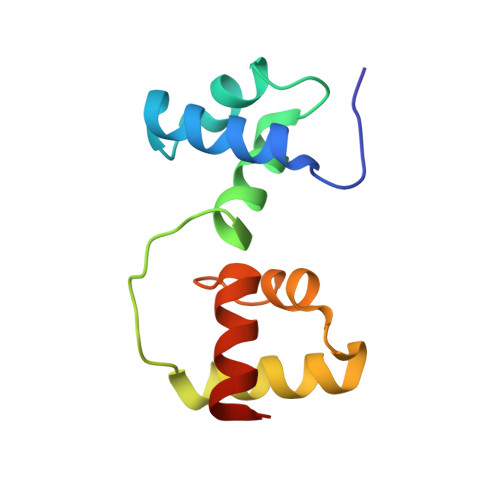 | |
UniProt & NIH Common Fund Data Resources | |||||
Find proteins for P06876 (Mus musculus) Explore P06876 Go to UniProtKB: P06876 | |||||
IMPC: MGI:97249 | |||||
Entity Groups | |||||
| Sequence Clusters | 30% Identity50% Identity70% Identity90% Identity95% Identity100% Identity | ||||
| UniProt Group | P06876 | ||||
Sequence AnnotationsExpand | |||||
| |||||
| Ligands 1 Unique | |||||
|---|---|---|---|---|---|
| ID | Chains | Name / Formula / InChI Key | 2D Diagram | 3D Interactions | |
| NA Query on NA | B [auth A], C [auth A] | SODIUM ION Na FKNQFGJONOIPTF-UHFFFAOYSA-N |  | ||
| Length ( Å ) | Angle ( ˚ ) |
|---|---|
| a = 38.722 | α = 90 |
| b = 28.25 | β = 90.87 |
| c = 44.129 | γ = 90 |
| Software Name | Purpose |
|---|---|
| CNS | refinement |
| DENZO | data reduction |
| SCALEPACK | data scaling |
| X-PLOR | phasing |
RCSB PDB (citation) is hosted by
RCSB PDB is a member of the

