Structural evidence for a possible role of reversible disulphide bridge formation in the elasticity of the muscle protein titin.
Mayans, O., Wuerges, J., Canela, S., Gautel, M., Wilmanns, M.(2001) Structure 9: 331-340
- PubMed: 11525170
- DOI: https://doi.org/10.1016/s0969-2126(01)00591-3
- Primary Citation of Related Structures:
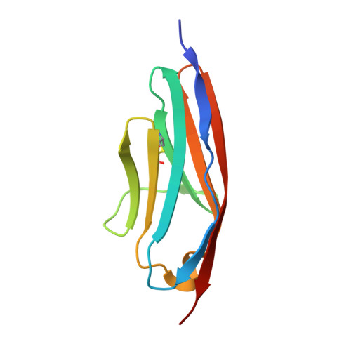
1G1C - PubMed Abstract:
The giant muscle protein titin contributes to the filament system in skeletal and cardiac muscle cells by connecting the Z disk and the central M line of the sarcomere. One of the physiological functions of titin is to act as a passive spring in the sarcomere, which is achieved by the elastic properties of its central I band region. Titin contains about 300 domains of which more than half are folded as immunoglobulin-like (Ig) domains. Ig domain segments of the I band of titin have been extensively used as templates to investigate the molecular basis of protein elasticity. The structure of the Ig domain I1 from the I band of titin has been determined to 2.1 A resolution. It reveals a novel, reversible disulphide bridge, which is neither required for correct folding nor changes the chemical stability of I1, but it is predicted to contribute mechanically to the elastic properties of titin in active sarcomeres. From the 92 Ig domains in the longest isoform of titin, at least 40 domains have a potential for disulphide bridge formation. We propose a model where the formation of disulphide bridges under oxidative stress conditions could regulate the elasticity of the I band in titin by increasing sarcomeric resistance. In this model, the formation of the disulphide bridge could refrain a possible directed motion of the two beta sheets or other mechanically stable entities of the I1 Ig domain with respect to each other when exposed to mechanical forces.
Organizational Affiliation:
EMBL Hamburg Outstation, Germany.