Energetic, structural, and antimicrobial analyses of beta-lactam side chain recognition by beta-lactamases.
Caselli, E., Powers, R.A., Blasczcak, L.C., Wu, C.Y., Prati, F., Shoichet, B.K.(2001) Chem Biol 8: 17-31
- PubMed: 11182316
- DOI: https://doi.org/10.1016/s1074-5521(00)00052-1
- Primary Citation of Related Structures:
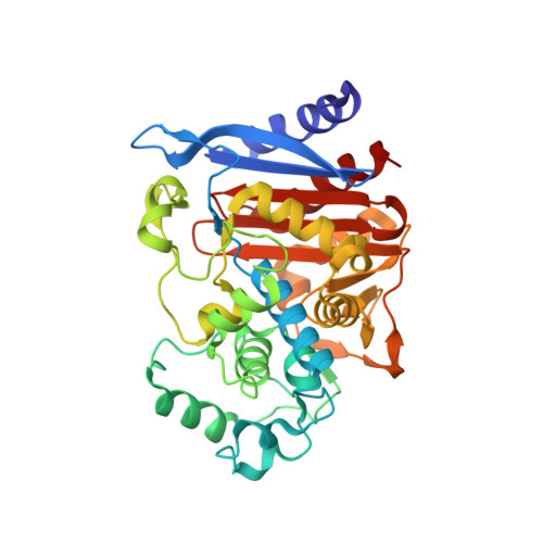
1FSW, 1FSY - PubMed Abstract:
Penicillins and cephalosporins are among the most widely used and successful antibiotics. The emergence of resistance to these beta-lactams, most often through bacterial expression of beta-lactamases, threatens public health. To understand how beta-lactamases recognize their substrates, it would be helpful to know their binding energies. Unfortunately, these have been difficult to measure because beta-lactams form covalent adducts with beta-lactamases. This has complicated functional analyses and inhibitor design. To investigate the contribution to interaction energy of the key amide (R1) side chain of beta-lactam antibiotics, eight acylglycineboronic acids that bear the side chains of characteristic penicillins and cephalosporins, as well as four other analogs, were synthesized. These transition-state analogs form reversible adducts with serine beta-lactamases. Therefore, binding energies can be calculated directly from K(i) values. The K(i) values measured span four orders of magnitude against the Group I beta-lactamase AmpC and three orders of magnitude against the Group II beta-lactamase TEM-1. The acylglycineboronic acids have K(i) values as low as 20 nM against AmpC and as low as 390 nM against TEM-1. The inhibitors showed little activity against serine proteases, such as chymotrypsin. R1 side chains characteristic of beta-lactam inhibitors did not have better affinity for AmpC than did side chains characteristic of beta-lactam substrates. Two of the inhibitors reversed the resistance of pathogenic bacteria to beta-lactams in cell culture. Structures of two inhibitors in their complexes with AmpC were determined by X-ray crystallography to 1.90 A and 1.75 A resolution; these structures suggest interactions that are important to the affinity of the inhibitors. Acylglycineboronic acids allow us to begin to dissect interaction energies between beta-lactam side chains and beta-lactamases. Surprisingly, there is little correlation between the affinity contributed by R1 side chains and their occurrence in beta-lactam inhibitors or beta-lactam substrates of serine beta-lactamases. Nevertheless, presented in acylglycineboronic acids, these side chains can lead to inhibitors with high affinities and specificities. The structures of their complexes with AmpC give a molecular context to their affinities and may guide the design of anti-resistance compounds in this series.
Organizational Affiliation:
Department of Molecular Pharmacology and Biological Chemistry, Northwestern University, Chicago, IL 60611, USA.