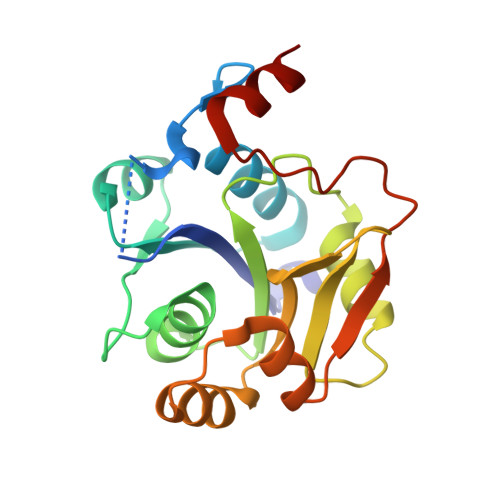
The crystal structure of the Escherichia coli MobA protein provides insight into molybdopterin guanine dinucleotide biosynthesis.
Lake, M.W., Temple, C.A., Rajagopalan, K.V., Schindelin, H.(2000) J Biol Chem 275: 40211-40217
- PubMed: 10978347
- DOI: https://doi.org/10.1074/jbc.M007406200
- Primary Citation of Related Structures:
1FR9, 1FRW - PubMed Abstract:
The molybdenum cofactor (Moco) is found in a variety of enzymes present in all phyla and comprises a family of related molecules containing molybdopterin (MPT), a tricyclic pyranopterin with a cis-dithiolene group, as the invariant essential moiety. MPT biosynthesis involves a conserved pathway, but some organisms perform additional reactions that modify MPT. In eubacteria, the cofactor is often present in a dinucleotide form combining MPT and a purine or pyrimidine nucleotide via a pyrophosphate linkage. In Escherichia coli, the MobA protein links a guanosine 5'-phosphate to MPT forming molybdopterin guanine dinucleotide. This reaction requires GTP, MgCl(2), and the MPT form of the cofactor and can efficiently reconstitute Rhodobacter sphaeroides apo-DMSOR, an enzyme that requires molybdopterin guanine dinucleotide for activity. In this paper, we present the crystal structure of MobA, a protein containing 194 amino acids. The MobA monomer has an alpha/beta architecture in which the N-terminal half of the molecule adopts a Rossman fold. The structure of MobA has striking similarity to Bacillus subtilis SpsA, a nucleotide-diphospho-sugar transferase involved in sporulation. The cocrystal structure of MobA and GTP reveals that the GTP-binding site is located in the N-terminal half of the molecule. Conserved residues located primarily in three signature sequence motifs form crucial interactions with the bound nucleotide. The binding site for MPT is located adjacent to the GTP-binding site in the C-terminal half of the molecule, which contains another set of conserved residues presumably involved in MPT binding.
Organizational Affiliation:
Department of Biochemistry and Center for Structural Biology, State University of New York at Stony Brook, Stony Brook, New York 11794-5215, USA.