1FEW

CRYSTAL STRUCTURE OF SMAC/DIABLO


Experimental Data Snapshot

  • Method: X-RAY DIFFRACTION
  • Resolution: 2.20 Å
  • R-Value Free: 0.259 
  • R-Value Work: 0.245 
  • R-Value Observed: 0.247 

wwPDB Validation   3D Report Full Report


This is version 1.3 of the entry. See complete history


Literature

Structural and biochemical basis of apoptotic activation by Smac/DIABLO.

Chai, J.Du, C.Wu, J.W.Kyin, S.Wang, X.Shi, Y.

(2000) Nature 406: 855-862

  • DOI: https://doi.org/10.1038/35022514
  • Primary Citation of Related Structures:  
    1FEW

  • PubMed Abstract: 

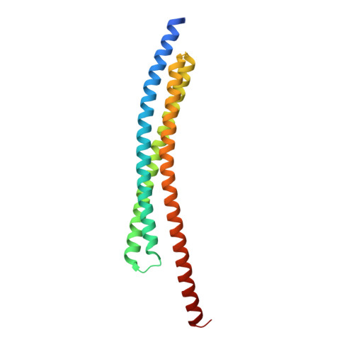
    Apoptosis (programmed cell death), an essential process in the development and homeostasis of metazoans, is carried out by caspases. The mitochondrial protein Smac/DIABLO performs a critical function in apoptosis by eliminating the inhibitory effect of IAPs (inhibitor of apoptosis proteins) on caspases. Here we show that Smac/DIABLO promotes not only the proteolytic activation of procaspase-3 but also the enzymatic activity of mature caspase-3, both of which depend upon its ability to interact physically with IAPs. The crystal structure of Smac/DIABLO at 2.2 A resolution reveals that it homodimerizes through an extensive hydrophobic interface. Missense mutations inactivating this dimeric interface significantly compromise the function of Smac/DIABLO. As in the Drosophila proteins Reaper, Grim and Hid, the amino-terminal amino acids of Smac/DIABLO are indispensable for its function, and a seven-residue peptide derived from the amino terminus promotes procaspase-3 activation in vitro. These results establish an evolutionarily conserved structural and biochemical basis for the activation of apoptosis by Smac/DIABLO.


  • Organizational Affiliation

    Department of Molecular Biology, Princeton University, New Jersey 08544, USA.


Macromolecules
Find similar proteins by:  (by identity cutoff)  |  3D Structure
Entity ID: 1
MoleculeChains Sequence LengthOrganismDetailsImage
SECOND MITOCHONDRIA-DERIVED ACTIVATOR OF CASPASES184Homo sapiensMutation(s): 0 
UniProt & NIH Common Fund Data Resources
Find proteins for Q9NR28 (Homo sapiens)
Explore Q9NR28 
Go to UniProtKB:  Q9NR28
PHAROS:  Q9NR28
GTEx:  ENSG00000184047 
Entity Groups  
Sequence Clusters30% Identity50% Identity70% Identity90% Identity95% Identity100% Identity
UniProt GroupQ9NR28
Sequence Annotations
Expand
  • Reference Sequence
Experimental Data & Validation

Experimental Data

  • Method: X-RAY DIFFRACTION
  • Resolution: 2.20 Å
  • R-Value Free: 0.259 
  • R-Value Work: 0.245 
  • R-Value Observed: 0.247 
  • Space Group: P 61 2 2
Unit Cell:
Length ( Å )Angle ( ˚ )
a = 108.5α = 90
b = 108.5β = 90
c = 70.4γ = 120
Software Package:
Software NamePurpose
DMmodel building
CNSrefinement
DENZOdata reduction
SCALEPACKdata scaling
DMphasing

Structure Validation

View Full Validation Report


Entry History 

Deposition Data

Revision History  (Full details and data files)

  • Version 1.0: 2000-09-13
    Type: Initial release
  • Version 1.1: 2008-04-27
    Changes: Version format compliance
  • Version 1.2: 2011-07-13
    Changes: Derived calculations, Version format compliance
  • Version 1.3: 2024-02-07
    Changes: Data collection, Database references