1FAW

GRAYLAG GOOSE HEMOGLOBIN (OXY FORM)


Experimental Data Snapshot

  • Method: X-RAY DIFFRACTION
  • Resolution: 3.09 Å
  • R-Value Free: 0.220 
  • R-Value Work: 0.176 
  • R-Value Observed: 0.176 

wwPDB Validation   3D Report Full Report


Ligand Structure Quality Assessment 


This is version 1.3 of the entry. See complete history


Literature

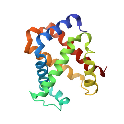
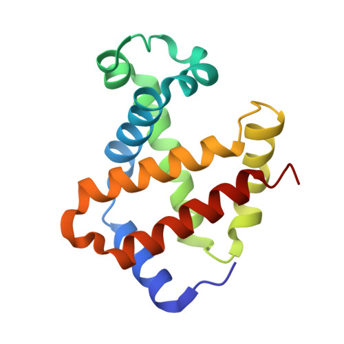
The structure of greylag goose oxy haemoglobin: the roles of four mutations compared with bar-headed goose haemoglobin.

Liang, Y.H.Liu, X.Z.Liu, S.H.Lu, G.Y.

(2001) Acta Crystallogr D Biol Crystallogr 57: 1850-1856

  • DOI: https://doi.org/10.1107/s0907444901016493
  • Primary Citation of Related Structures:  
    1FAW

  • PubMed Abstract: 

    The greylag goose (Anser anser), which lives on lowlands and cannot tolerate hypoxic conditions, presents a striking contrast to its close relative the bar-headed goose (A. indicus), which lives at high altitude and possesses high-altitude hypoxia adaptation. There are only four amino-acid residue differences at alpha18, alpha63, alpha119 and beta125 between the haemoglobins of the two species. The crystal structure of greylag goose oxy haemoglobin was determined at 3.09 A resolution. Its quaternary structure is slightly different from that of the bar-headed goose oxy haemoglobin, with a rotation of 2.8 degrees in relative orientation of the two dimers. Of the four mutations, those at alpha119 and beta125 produce contact changes in the alpha(1)beta(1) interface and may be responsible for the differences in intrinsic oxygen affinity between the two species; those at alpha18 and alpha63 may be responsible for the differences in quaternary structure between the two species.


  • Organizational Affiliation

    National Laboratory of Protein Engineering and Plant Genetic Engineering, College of Life Sciences, Peking University, Beijing 100871, People's Repbulic of China.


Macromolecules
Find similar proteins by:  (by identity cutoff)  |  3D Structure
Entity ID: 1
MoleculeChains Sequence LengthOrganismDetailsImage
HEMOGLOBIN (ALPHA SUBUNIT)
A, C
141Anser anserMutation(s): 0 
UniProt
Find proteins for P01989 (Anser anser anser)
Explore P01989 
Go to UniProtKB:  P01989
Entity Groups  
Sequence Clusters30% Identity50% Identity70% Identity90% Identity95% Identity100% Identity
UniProt GroupP01989
Sequence Annotations
Expand
  • Reference Sequence
Find similar proteins by:  (by identity cutoff)  |  3D Structure
Entity ID: 2
MoleculeChains Sequence LengthOrganismDetailsImage
HEMOGLOBIN (BETA SUBUNIT)
B, D
146Anser anserMutation(s): 0 
UniProt
Find proteins for P02117 (Anser anser anser)
Explore P02117 
Go to UniProtKB:  P02117
Entity Groups  
Sequence Clusters30% Identity50% Identity70% Identity90% Identity95% Identity100% Identity
UniProt GroupP02117
Sequence Annotations
Expand
  • Reference Sequence
Experimental Data & Validation

Experimental Data

  • Method: X-RAY DIFFRACTION
  • Resolution: 3.09 Å
  • R-Value Free: 0.220 
  • R-Value Work: 0.176 
  • R-Value Observed: 0.176 
  • Space Group: P 1 21 1
Unit Cell:
Length ( Å )Angle ( ˚ )
a = 57.552α = 90
b = 80.624β = 102.75
c = 72.571γ = 90
Software Package:
Software NamePurpose
CNSrefinement
DENZOdata reduction
SCALEPACKdata scaling
CNSphasing

Structure Validation

View Full Validation Report


Ligand Structure Quality Assessment 


Entry History 

Deposition Data

Revision History  (Full details and data files)

  • Version 1.0: 2001-12-05
    Type: Initial release
  • Version 1.1: 2008-04-27
    Changes: Version format compliance
  • Version 1.2: 2011-07-13
    Changes: Version format compliance
  • Version 1.3: 2024-02-07
    Changes: Data collection, Database references, Derived calculations