1F1C

CRYSTAL STRUCTURE OF CYTOCHROME C549


Experimental Data Snapshot

  • Method: X-RAY DIFFRACTION
  • Resolution: 2.30 Å
  • R-Value Free: 0.260 
  • R-Value Work: 0.217 
  • R-Value Observed: 0.237 

wwPDB Validation   3D Report Full Report


Ligand Structure Quality Assessment 


This is version 2.0 of the entry. See complete history


Literature

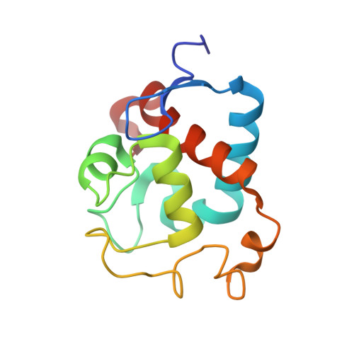
Structures of cytochrome c-549 and cytochrome c6 from the cyanobacterium Arthrospira maxima.

Sawaya, M.R.Krogmann, D.W.Serag, A.Ho, K.K.Yeates, T.O.Kerfeld, C.A.

(2001) Biochemistry 40: 9215-9225

  • DOI: https://doi.org/10.1021/bi002679p
  • Primary Citation of Related Structures:  
    1F1C, 1F1F

  • PubMed Abstract: 

    Cytochrome c(6) and cytochrome c-549 are small (89 and 130 amino acids, respectively) monoheme cytochromes that function in photosynthesis. They appear to have descended relatively recently from the same ancestral gene but have diverged to carry out very different functional roles, underscored by the large difference between their midpoint potentials of nearly 600 mV. We have determined the X-ray crystal structures of both proteins isolated from the cyanobacterium Arthrospira maxima. The two structures are remarkably similar, superimposing on backbone atoms with an rmsd of 0.7 A. Comparison of the two structures suggests that differences in solvent exposure of the heme and the electrostatic environment of the heme propionates, as well as in heme iron ligation, are the main determinants of midpoint potential in the two proteins. In addition, the crystal packing of both A. maxima cytochrome c-549 and cytochrome c(6) suggests that the proteins oligomerize. Finally, the cytochrome c-549 dimer we observe can be readily fit into the recently described model of cyanobacterial photosystem II.


  • Organizational Affiliation

    Molecular Biology Institute, University of California, Los Angeles, Box 951570, Los Angeles, California 90095-1570, USA.


Macromolecules
Find similar proteins by:  (by identity cutoff)  |  3D Structure
Entity ID: 1
MoleculeChains Sequence LengthOrganismDetailsImage
CYTOCHROME C549
A, B
129Limnospira maximaMutation(s): 0 
UniProt
Find proteins for P82603 (Limnospira maxima)
Explore P82603 
Go to UniProtKB:  P82603
Entity Groups  
Sequence Clusters30% Identity50% Identity70% Identity90% Identity95% Identity100% Identity
UniProt GroupP82603
Sequence Annotations
Expand
  • Reference Sequence
Experimental Data & Validation

Experimental Data

  • Method: X-RAY DIFFRACTION
  • Resolution: 2.30 Å
  • R-Value Free: 0.260 
  • R-Value Work: 0.217 
  • R-Value Observed: 0.237 
  • Space Group: P 1 21 1
Unit Cell:
Length ( Å )Angle ( ˚ )
a = 36.5α = 90
b = 84.4β = 96.5
c = 43.8γ = 90
Software Package:
Software NamePurpose
EPMRphasing
CNSrefinement
DENZOdata reduction
SCALEPACKdata scaling

Structure Validation

View Full Validation Report


Ligand Structure Quality Assessment 


Entry History 

Deposition Data

Revision History  (Full details and data files)

  • Version 1.0: 2001-08-08
    Type: Initial release
  • Version 1.1: 2008-04-27
    Changes: Version format compliance
  • Version 1.2: 2011-07-13
    Changes: Version format compliance
  • Version 2.0: 2021-03-03
    Changes: Advisory, Atomic model, Data collection, Derived calculations, Non-polymer description, Structure summary