1EYT

CRYSTAL STRUCTURE OF HIGH-POTENTIAL IRON-SULFUR PROTEIN FROM THERMOCHROMATIUM TEPIDUM


Experimental Data Snapshot

  • Method: X-RAY DIFFRACTION
  • Resolution: 1.50 Å
  • R-Value Free: 0.238 
  • R-Value Work: 0.212 
  • R-Value Observed: 0.213 

wwPDB Validation   3D Report Full Report


This is version 1.3 of the entry. See complete history


Literature

Crystal structures of photosynthetic reaction center and high-potential iron-sulfur protein from Thermochromatium tepidum: thermostability and electron transfer.

Nogi, T.Fathir, I.Kobayashi, M.Nozawa, T.Miki, K.

(2000) Proc Natl Acad Sci U S A 97: 13561-13566

  • DOI: https://doi.org/10.1073/pnas.240224997
  • Primary Citation of Related Structures:  
    1EYS, 1EYT

  • PubMed Abstract: 

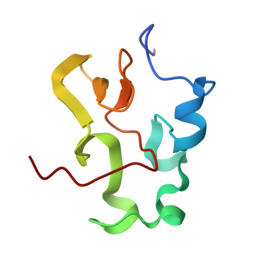
    The reaction center (RC) of photosynthetic bacteria is a membrane protein complex that promotes a light-induced charge separation during the primary process of photosynthesis. In the photosynthetic electron transfer chain, the soluble electron carrier proteins transport electrons to the RC and reduce the photo-oxidized special-pair of bacteriochlorophyll. The high-potential iron-sulfur protein (HiPIP) is known to serve as an electron donor to the RC in some species, where the c-type cytochrome subunit, the peripheral subunit of the RC, directly accepts electrons from the HiPIP. Here we report the crystal structures of the RC and the HiPIP from Thermochromatium (Tch.) tepidum, at 2.2-A and 1.5-A resolution, respectively. Tch. tepidum can grow at the highest temperature of all known purple bacteria, and the Tch. tepidum RC shows some degree of stability to high temperature. Comparison with the RCs of mesophiles, such as Blastochloris viridis, has shown that the Tch. tepidum RC possesses more Arg residues at the membrane surface, which might contribute to the stability of this membrane protein. The RC and the HiPIP both possess hydrophobic patches on their respective surfaces, and the HiPIP is expected to interact with the cytochrome subunit by hydrophobic interactions near the heme-1, the most distal heme to the special-pair.


  • Organizational Affiliation

    Department of Chemistry, Graduate School of Science, Kyoto University, Sakyo-ku, Kyoto 606-8502, Japan.


Macromolecules
Find similar proteins by:  (by identity cutoff)  |  3D Structure
Entity ID: 1
MoleculeChains Sequence LengthOrganismDetailsImage
HIGH-POTENTIAL IRON-SULFUR PROTEIN83Thermochromatium tepidumMutation(s): 0 
UniProt
Find proteins for P80176 (Thermochromatium tepidum)
Explore P80176 
Go to UniProtKB:  P80176
Entity Groups  
Sequence Clusters30% Identity50% Identity70% Identity90% Identity95% Identity100% Identity
UniProt GroupP80176
Sequence Annotations
Expand
  • Reference Sequence
Small Molecules
Ligands 1 Unique
IDChains Name / Formula / InChI Key2D Diagram3D Interactions
SF4
Query on SF4

Download Ideal Coordinates CCD File 
B [auth A]IRON/SULFUR CLUSTER
Fe4 S4
LJBDFODJNLIPKO-UHFFFAOYSA-N
Experimental Data & Validation

Experimental Data

  • Method: X-RAY DIFFRACTION
  • Resolution: 1.50 Å
  • R-Value Free: 0.238 
  • R-Value Work: 0.212 
  • R-Value Observed: 0.213 
  • Space Group: P 21 21 21
Unit Cell:
Length ( Å )Angle ( ˚ )
a = 47.118α = 90
b = 59.592β = 90
c = 23.62γ = 90
Software Package:
Software NamePurpose
AMoREphasing
X-PLORrefinement
DENZOdata reduction
SCALEPACKdata scaling

Structure Validation

View Full Validation Report


Entry History 

Deposition Data

Revision History  (Full details and data files)

  • Version 1.0: 2000-12-13
    Type: Initial release
  • Version 1.1: 2008-04-27
    Changes: Version format compliance
  • Version 1.2: 2011-07-13
    Changes: Version format compliance
  • Version 1.3: 2024-02-07
    Changes: Data collection, Database references, Derived calculations