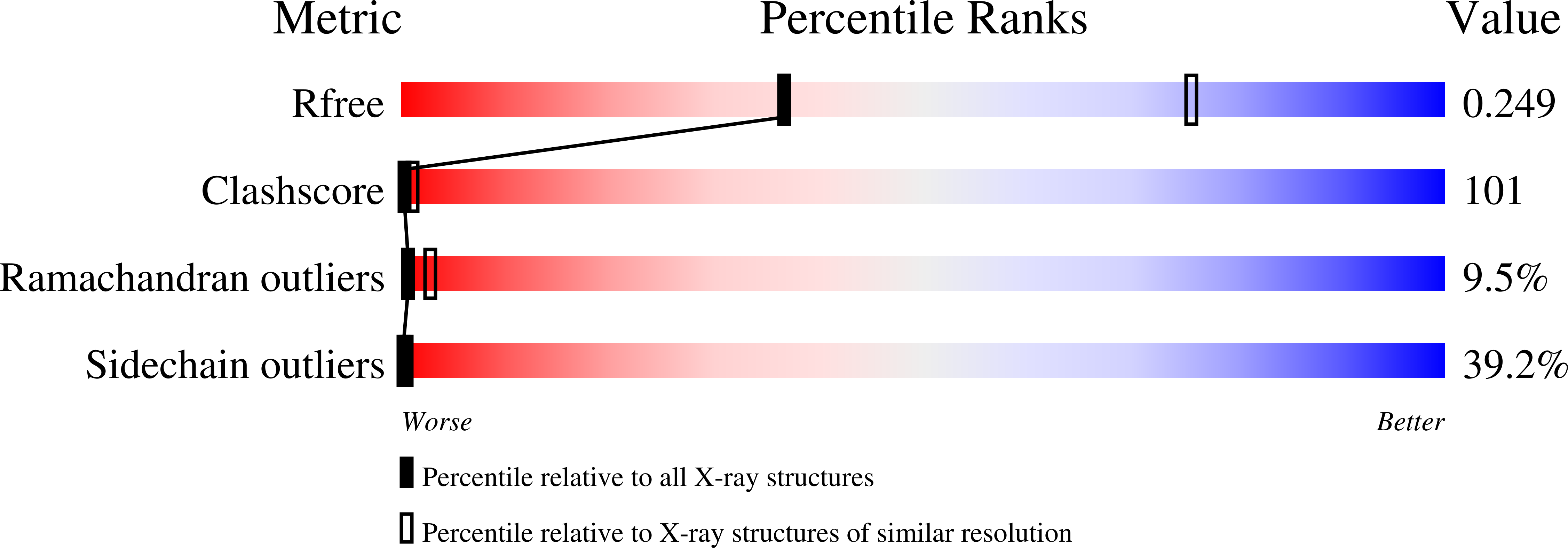
Crystal structure of glycosyltrehalose trehalohydrolase from the hyperthermophilic archaeum Sulfolobus solfataricus.
Feese, M.D., Kato, Y., Tamada, T., Kato, M., Komeda, T., Miura, Y., Hirose, M., Hondo, K., Kobayashi, K., Kuroki, R.(2000) J Mol Biol 301: 451-464
- PubMed: 10926520
- DOI: https://doi.org/10.1006/jmbi.2000.3977
- Primary Citation of Related Structures:
1EH9, 1EHA - PubMed Abstract:
The crystal structure of glycosyltrehalose trehalohydrolase from the hyperthermophilic archaeum Sulfolobus solfataricus KM1 has been solved by multiple isomorphous replacement. The enzyme is an alpha-amylase (family 13) with unique exo-amylolytic activity for glycosyltrehalosides. It cleaves the alpha-1,4 glycosidic bond adjacent to the trehalose moiety to release trehalose and maltooligo saccharide. Unlike most other family 13 glycosidases, the enzyme does not require Ca(2+) for activity, and it contains an N-terminal extension of approximately 100 amino acid residues that is homologous to N-terminal domains found in many glycosidases that recognize branched oligosaccharides. Crystallography revealed the enzyme to exist as a homodimer covalently linked by an intermolecular disulfide bond at residue C298. The existence of the intermolecular disulfide bond was confirmed by biochemical analysis and mutagenesis. The N-terminal extension forms an independent domain connected to the catalytic domain by an extended linker. The functionally essential Ca(2+) binding site found in the B domain of alpha-amylases and many other family 13 glycosidases was found to be replaced by hydrophobic packing interactions. The enzyme also contains a very unusual excursion in the (beta/alpha)(8) barrel structure of the catalytic domain. This excursion originates from the bottom of the (beta/alpha)(8) barrel between helix 6 and strand 7, but folds upward in a distorted alpha-hairpin structure to form a part of the substrate binding cleft wall that is possibly critical for the enzyme's unique substrate selectivity. Participation of an alpha-beta loop in the formation of the substrate binding cleft is a novel feature that is not observed in other known (beta/alpha)(8) enzymes.
Organizational Affiliation:
Central Laboratories for Key Technology, Kirin Brewery Co. Ltd, 1-13-5 Fukuura, Kanazawa, Yokohama 236, Japan,


