Structure and binding specificity of the second N-terminal cellulose-binding domain from Cellulomonas fimi endoglucanase C.
Brun, E., Johnson, P.E., Creagh, A.L., Tomme, P., Webster, P., Haynes, C.A., McIntosh, L.P.(2000) Biochemistry 39: 2445-2458
- PubMed: 10704194
- DOI: https://doi.org/10.1021/bi992079u
- Primary Citation of Related Structures:
1CX1 - PubMed Abstract:
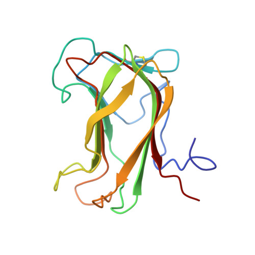
The 1,4-beta-glucanase CenC from Cellulomonas fimi contains two cellulose-binding domains, CBD(N1) and CBD(N2), arranged in tandem at its N-terminus. These homologous CBDs are distinct in their selectivity for binding amorphous and not crystalline cellulose. Multidimensional heteronuclear nuclear magnetic resonance (NMR) spectroscopy was used to determine the tertiary structure of CBD(N2) in the presence of saturating amounts of cellopentaose. A total of 1996 experimental restraints were used to calculate an ensemble of 21 final structures for the protein. CBD(Nu2) is composed of 11 beta-strands, folded into two antiparallel beta-sheets, with a topology of a jellyroll beta-sandwich. On the basis of patterns of chemical shift perturbations accompanying the addition of cellooligosaccharides, as well as the observation of intermolecular protein-sugar NOE interactions, the cellulose-binding site of CBD(N2) was identified as a cleft that lies across one face of the beta-sandwich. The thermodynamic basis for the binding of cellooligosaccharides was investigated using isothermal titration calorimetry and NMR spectroscopy. Binding is enthalpically driven and consistent with a structural model involving hydrogen bonding between the equatorial hydroxyls of the glucopyranosyl rings and polar amino acid side chains lining the CBD(N2) cleft. Affinity electrophoresis was used to determine that CBD(N2) also binds soluble beta-1,4-linked polymers of glucose, including hydroxyethylcellulose and beta-1,3-1,4-glucans. This study complements a previous analysis of CBD(N1) [Johnson, P. E., Joshi, M. D., Tomme, P., Kilburn, D. G., and McIntosh, L. P. (1996) Biochemistry 35, 14381-14394] and demonstrates that the homologous CBDs from CenC share very similar structures and sugar binding properties.
Organizational Affiliation:
Protein Engineering Network of Centres of Excellence, Department of Biochemistry and Molecular Biology, The University of British Columbia, Vancouver, Canada.