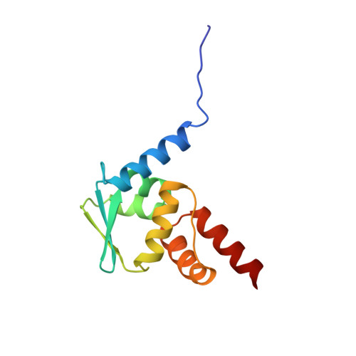
Crystal structure of the BTB domain from PLZF.
Ahmad, K.F., Engel, C.K., Prive, G.G.(1998) Proc Natl Acad Sci U S A 95: 12123-12128
- PubMed: 9770450
- DOI: https://doi.org/10.1073/pnas.95.21.12123
- Primary Citation of Related Structures:
1BUO - PubMed Abstract:
The BTB domain (also known as the POZ domain) is an evolutionarily conserved protein-protein interaction motif found at the N terminus of 5-10% of C2H2-type zinc-finger transcription factors, as well as in some actin-associated proteins bearing the kelch motif. Many BTB proteins are transcriptional regulators that mediate gene expression through the control of chromatin conformation. In the human promyelocytic leukemia zinc finger (PLZF) protein, the BTB domain has transcriptional repression activity, directs the protein to a nuclear punctate pattern, and interacts with components of the histone deacetylase complex. The association of the PLZF BTB domain with the histone deacetylase complex provides a mechanism of linking the transcription factor with enzymatic activities that regulate chromatin conformation. The crystal structure of the BTB domain of PLZF was determined at 1.9 A resolution and reveals a tightly intertwined dimer with an extensive hydrophobic interface. Approximately one-quarter of the monomer surface area is involved in the dimer intermolecular contact. These features are typical of obligate homodimers, and we expect the full-length PLZF protein to exist as a branched transcription factor with two C-terminal DNA-binding regions. A surface-exposed groove lined with conserved amino acids is formed at the dimer interface, suggestive of a peptide-binding site. This groove may represent the site of interaction of the PLZF BTB domain with nuclear corepressors or other nuclear proteins.
Organizational Affiliation:
Division of Molecular and Structural Biology, Ontario Cancer Institute, and the Department of Medical Biophysics, University of Toronto, 610 University Avenue, Toronto, Ontario, Canada M5G 2M9.