Function from structure? The crystal structure of human phosphatidylethanolamine-binding protein suggests a role in membrane signal transduction.
Banfield, M.J., Barker, J.J., Perry, A.C., Brady, R.L.(1998) Structure 6: 1245-1254
- PubMed: 9782050
- DOI: https://doi.org/10.1016/s0969-2126(98)00125-7
- Primary Citation of Related Structures:
1BD9, 1BEH - PubMed Abstract:
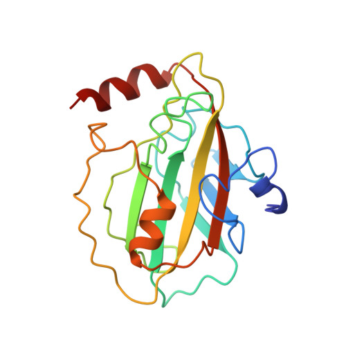
Proteins belonging to the phosphatidylethanolamine-binding protein (PEBP) family are highly conserved throughout nature and have no significant sequence homology with other proteins of known structure or function. A variety of biological roles have previously been described for members of this family, including lipid binding, roles as odorant effector molecules or opioids, interaction with the cell-signalling machinery, regulation of flowering plant stem architecture, and a function as a precursor protein of a bioactive brain neuropeptide. To date, no experimentally derived structural information has been available for this protein family. In this study we have used X-ray crystallography to determine the three-dimensional structure of human PEBP (hPEBP), in an attempt to clarify the biological role of this unique protein family. The crystal structures of two forms of hPEBP have been determined: one in the native state (at 2.05 A resolution) and one in complex with cacodylate (at 1.75 A resolution). The crystal structures reveal that hPEBP adopts a novel protein topology, dominated by the presence of a large central beta sheet, and is expected to represent the archaetypal fold for this family of proteins. Two potential functional sites have been identified from the structure: a putative ligand-binding site and a coupled cleavage site. hPEBP forms a dimer in the crystal with a distinctive dipole moment that may orient the oligomer for membrane binding. The crystal structure of hPEBP suggests that the ligand-binding site could accommodate the phosphate head groups of membrane lipids, therefore allowing the protein to adhere to the inner leaf of bilipid membranes where it would be ideally positioned to relay signals from the membrane to the cytoplasm. The structure also suggests that ligand binding may lead to coordinated release of the N-terminal region of the protein to form the hippocampal neurostimulatory peptide, which is known to be active in the development of the hippocampus. These studies are consistent with a primary biological role for hPEBP as a transducer of signals from the interior membrane surface.
Organizational Affiliation:
Department of Biochemistry University of Bristol Bristol, BS8 1TD, UK. M.Banfield@bristol.ac.uk