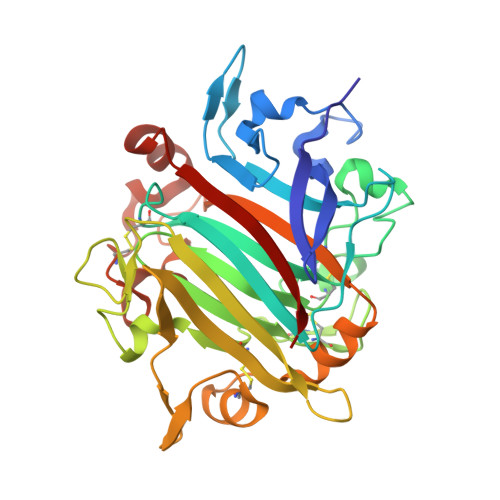
The crystal structure of RsSymEG1 reveals a unique form of smaller GH7 endoglucanases alongside GH7 cellobiohydrolases in protist symbionts of termites.
Haataja, T., Hansson, H., Moriya, S., Sandgren, M., Stahlberg, J.(2024) FEBS J 291: 1168-1185
- PubMed: 38073120
- DOI: https://doi.org/10.1111/febs.17029
- Primary Citation of Related Structures:
8POF - PubMed Abstract:
Glycoside hydrolase family 7 (GH7) cellulases are key enzymes responsible for carbon cycling on earth through their role in cellulose degradation and constitute highly important industrial enzymes as well. Although these enzymes are found in a wide variety of evolutionarily distant organisms across eukaryotes, they exhibit remarkably conserved features within two groups: exo-acting cellobiohydrolases and endoglucanases. However, recently reports have emerged of a separate clade of GH7 endoglucanases from protist symbionts of termites that are 60-80 amino acids shorter. In this work, we describe the first crystal structure of a short GH7 endoglucanase, RsSymEG1, from a symbiont of the lower termite Reticulitermes speratus. A more open flat surface and shorter loops around the non-reducing end of the cellulose-binding cleft indicate enhanced access to cellulose chains on the surface of cellulose microfibrils. Additionally, when comparing activities on polysaccharides to a typical fungal GH7 endoglucanase (Trichoderma longibrachiatum Cel7B), RsSymEG1 showed significantly faster initial hydrolytic activity. We also examine the prevalence and diversity of GH7 enzymes that the symbionts provide to the termite host, compare overall structures and substrate binding between cellobiohydrolase and long and short endoglucanase, and highlight the presence of similar short GH7s in other organisms.
Organizational Affiliation:
Department of Molecular Sciences, Swedish University of Agricultural Sciences, Uppsala, Sweden.