7QAZ

Prim-Pol Domain of CRISPR-associated Prim-Pol (CAPP) from Marinitoga sp. 1137 - Primer Initiation Complex


Experimental Data Snapshot

  • Method: X-RAY DIFFRACTION
  • Resolution: 2.11 Å
  • R-Value Free: 0.261 
  • R-Value Work: 0.210 
  • R-Value Observed: 0.212 

wwPDB Validation   3D Report Full Report


Ligand Structure Quality Assessment 


This is version 1.5 of the entry. See complete history


Literature

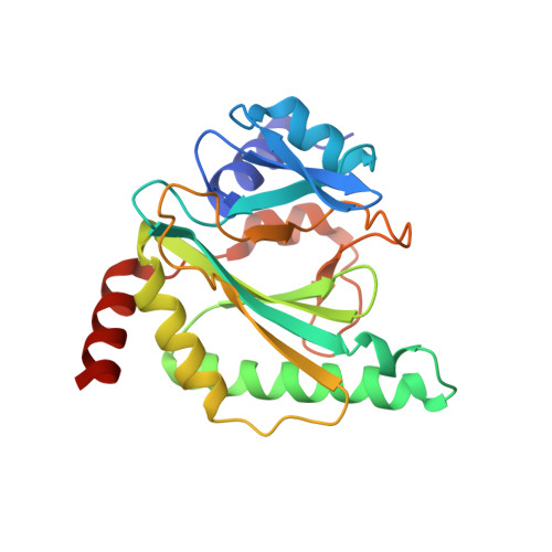
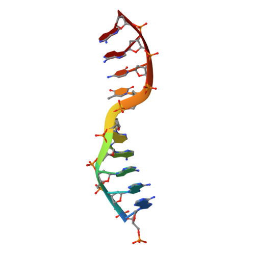
Molecular basis for the initiation of DNA primer synthesis.

Li, A.W.H.Zabrady, K.Bainbridge, L.J.Zabrady, M.Naseem-Khan, S.Berger, M.B.Kolesar, P.Cisneros, G.A.Doherty, A.J.

(2022) Nature 605: 767-773

  • DOI: https://doi.org/10.1038/s41586-022-04695-0
  • Primary Citation of Related Structures:  
    7NQD, 7NQE, 7NQF, 7P9J, 7QAZ

  • PubMed Abstract: 

    During the initiation of DNA replication, oligonucleotide primers are synthesized de novo by primases and are subsequently extended by replicative polymerases to complete genome duplication. The primase-polymerase (Prim-Pol) superfamily is a diverse grouping of primases, which includes replicative primases and CRISPR-associated primase-polymerases (CAPPs) involved in adaptive immunity 1-3 . Although much is known about the activities of these enzymes, the precise mechanism used by primases to initiate primer synthesis has not been elucidated. Here we identify the molecular bases for the initiation of primer synthesis by CAPP and show that this mechanism is also conserved in replicative primases. The crystal structure of a primer initiation complex reveals how the incoming nucleotides are positioned within the active site, adjacent to metal cofactors and paired to the templating single-stranded DNA strand, before synthesis of the first phosphodiester bond. Furthermore, the structure of a Prim-Pol complex with double-stranded DNA shows how the enzyme subsequently extends primers in a processive polymerase mode. The structural and mechanistic studies presented here establish how Prim-Pol proteins instigate primer synthesis, revealing the requisite molecular determinants for primer synthesis within the catalytic domain. This work also establishes that the catalytic domain of Prim-Pol enzymes, including replicative primases, is sufficient to catalyse primer formation.


  • Organizational Affiliation

    Genome Damage and Stability Centre, School of Life Sciences, University of Sussex, Brighton, UK.


Macromolecules

Find similar proteins by:  (by identity cutoff)  |  3D Structure
Entity ID: 1
MoleculeChains Sequence LengthOrganismDetailsImage
TPR_REGION domain-containing protein
A, B, C
219Marinitoga sp. 1137Mutation(s): 5 
Gene Names: Marpi_0402
UniProt
Find proteins for H2J4R1 (Marinitoga piezophila (strain DSM 14283 / JCM 11233 / KA3))
Explore H2J4R1 
Go to UniProtKB:  H2J4R1
Entity Groups  
Sequence Clusters30% Identity50% Identity70% Identity90% Identity95% Identity100% Identity
UniProt GroupH2J4R1
Sequence Annotations
Expand
  • Reference Sequence

Find similar nucleic acids by:  Sequence   |   3D Structure  

Entity ID: 2
MoleculeChains LengthOrganismImage
Templating strand9synthetic construct
Sequence Annotations
Expand
  • Reference Sequence
Small Molecules
Ligands 3 Unique
IDChains Name / Formula / InChI Key2D Diagram3D Interactions
GTP (Subject of Investigation/LOI)
Query on GTP

Download Ideal Coordinates CCD File 
K [auth B]GUANOSINE-5'-TRIPHOSPHATE
C10 H16 N5 O14 P3
XKMLYUALXHKNFT-UUOKFMHZSA-N
DZ4 (Subject of Investigation/LOI)
Query on DZ4

Download Ideal Coordinates CCD File 
F [auth A],
J [auth B],
L [auth B],
O [auth C]
2'-deoxy-5'-O-[(R)-hydroxy{[(R)-hydroxy(phosphonooxy)phosphoryl]amino}phosphoryl]adenosine
C10 H17 N6 O11 P3
WKIPJDSLGCBQCU-RRKCRQDMSA-N
CO (Subject of Investigation/LOI)
Query on CO

Download Ideal Coordinates CCD File 
E [auth A]
G [auth B]
H [auth B]
I [auth B]
M [auth C]
E [auth A],
G [auth B],
H [auth B],
I [auth B],
M [auth C],
N [auth C],
P [auth D]
COBALT (II) ION
Co
XLJKHNWPARRRJB-UHFFFAOYSA-N
Experimental Data & Validation

Experimental Data

  • Method: X-RAY DIFFRACTION
  • Resolution: 2.11 Å
  • R-Value Free: 0.261 
  • R-Value Work: 0.210 
  • R-Value Observed: 0.212 
  • Space Group: P 21 21 21
Unit Cell:
Length ( Å )Angle ( ˚ )
a = 227.91α = 90
b = 39.62β = 90
c = 79.77γ = 90
Software Package:
Software NamePurpose
xia2data reduction
PHENIXrefinement
XSCALEdata scaling
PHASERphasing
Cootmodel building

Structure Validation

View Full Validation Report


Ligand Structure Quality Assessment 


Entry History & Funding Information

Deposition Data


Funding OrganizationLocationGrant Number
Biotechnology and Biological Sciences Research Council (BBSRC)United KingdomBB/S008691/1
Biotechnology and Biological Sciences Research Council (BBSRC)United KingdomBB/M008800/1
Biotechnology and Biological Sciences Research Council (BBSRC)United KingdomBB/P007031/1

Revision History  (Full details and data files)

  • Version 1.0: 2022-05-04
    Type: Initial release
  • Version 1.1: 2022-05-18
    Changes: Database references
  • Version 1.2: 2022-05-25
    Changes: Database references
  • Version 1.3: 2022-06-08
    Changes: Database references
  • Version 1.4: 2022-11-02
    Changes: Derived calculations
  • Version 1.5: 2024-01-31
    Changes: Data collection, Refinement description