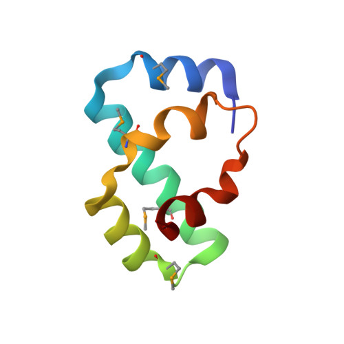
Structure and Function of the T4 Spackle Protein Gp61.3.
Kanamaru, S., Uchida, K., Nemoto, M., Fraser, A., Arisaka, F., Leiman, P.G.(2020) Viruses 12
- PubMed: 32987925
- DOI: https://doi.org/10.3390/v12101070
- Primary Citation of Related Structures:
7CN6, 7CN7 - PubMed Abstract:
The bacteriophage T4 genome contains two genes that code for proteins with lysozyme activity- e and 5 . Gene e encodes the well-known T4 lysozyme (commonly called T4L) that functions to break the peptidoglycan layer late in the infection cycle, which is required for liberating newly assembled phage progeny. Gene product 5 (gp5) is the tail-associated lysozyme, a component of the phage particle. It forms a spike at the tip of the tail tube and functions to pierce the outer membrane of the Escherichia coli host cell after the phage has attached to the cell surface. Gp5 contains a T4L-like lysozyme domain that locally digests the peptidoglycan layer upon infection. The T4 Spackle protein (encoded by gene 61.3 ) has been thought to play a role in the inhibition of gp5 lysozyme activity and, as a consequence, in making cells infected by bacteriophage T4 resistant to later infection by T4 and closely related phages. Here we show that (1) gp61.3 is secreted into the periplasm where its N-terminal periplasm-targeting peptide is cleaved off; (2) gp61.3 forms a 1:1 complex with the lysozyme domain of gp5 (gp5Lys); (3) gp61.3 selectively inhibits the activity of gp5, but not that of T4L; (4) overexpression of gp5 causes cell lysis. We also report a crystal structure of the gp61.3-gp5Lys complex that demonstrates that unlike other known lysozyme inhibitors, gp61.3 does not interact with the active site cleft. Instead, it forms a "wall" that blocks access of an extended polysaccharide substrate to the cleft and, possibly, locks the enzyme in an "open-jaw"-like conformation making catalysis impossible.
Organizational Affiliation:
Department of Life Science and Technology, Tokyo Institute of Technology, S2-7 4259 Nagatsuta-cho, Midori-ku, Yokohama, Kanagawa 226-8503, Japan.