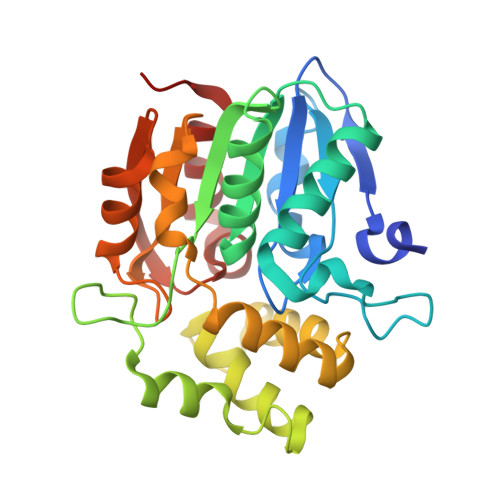
5DNU

Crystal structure of Striga KAI2-like protein in complex with karrikin


Experimental Data Snapshot

  • Method: X-RAY DIFFRACTION
  • Resolution: 1.20 Å
  • R-Value Free: 0.155 
  • R-Value Work: 0.140 
  • R-Value Observed: 0.141 

wwPDB Validation   3D Report Full Report


Ligand Structure Quality Assessment 


This is version 1.3 of the entry. See complete history


Literature

Structural basis of unique ligand specificity of KAI2-like protein from parasitic weed Striga hermonthica

Xu, Y.Miyakawa, T.Nakamura, H.Nakamura, A.Imamura, Y.Asami, T.Tanokura, M.

(2016) Sci Rep 6: 31386-31386

  • DOI: https://doi.org/10.1038/srep31386
  • Primary Citation of Related Structures:  
    5DNU, 5DNV, 5DNW

  • PubMed Abstract: 

    The perception of two plant germination inducers, karrikins and strigolactones, are mediated by the proteins KAI2 and D14. Recently, KAI2-type proteins from parasitic weeds, which are possibly related to seed germination induced by strigolactone, have been classified into three clades characterized by different responses to karrikin/strigolactone. Here we characterized a karrikin-binding protein in Striga (ShKAI2iB) that belongs to intermediate-evolving KAI2 and provided the structural bases for its karrikin-binding specificity. Binding assays showed that ShKAI2iB bound karrikins but not strigolactone, differing from other KAI2 and D14. The crystal structures of ShKAI2iB and ShKAI2iB-karrikin complex revealed obvious structural differences in a helix located at the entry of its ligand-binding cavity. This results in a smaller closed pocket, which is also the major cause of ShKAI2iB's specificity of binding karrikin. Our structural study also revealed that a few non-conserved amino acids led to the distinct ligand-binding profile of ShKAI2iB, suggesting that the evolution of KAI2 resulted in its diverse functions.


  • Organizational Affiliation

    Department of Applied Biological Chemistry, Graduate School of Agricultural and Life Sciences, The University of Tokyo, 1-1-1 Yayoi, Bunkyo-ku, Tokyo 113-8657, Japan.


Macromolecules
Find similar proteins by:  (by identity cutoff)  |  3D Structure
Entity ID: 1
MoleculeChains Sequence LengthOrganismDetailsImage
ShKAI2iB275Striga hermonthicaMutation(s): 0 
UniProt
Find proteins for A0A0M3PN85 (Striga hermonthica)
Explore A0A0M3PN85 
Go to UniProtKB:  A0A0M3PN85
Entity Groups  
Sequence Clusters30% Identity50% Identity70% Identity90% Identity95% Identity100% Identity
UniProt GroupA0A0M3PN85
Sequence Annotations
Expand
  • Reference Sequence
Small Molecules
Ligands 5 Unique
IDChains Name / Formula / InChI Key2D Diagram3D Interactions
KKN
Query on KKN

Download Ideal Coordinates CCD File 
B [auth A]3-methyl-2H-furo[2,3-c]pyran-2-one
C8 H6 O3
JUTMAMXOAOYKHT-UHFFFAOYSA-N
BEZ
Query on BEZ

Download Ideal Coordinates CCD File 
T [auth A]BENZOIC ACID
C7 H6 O2
WPYMKLBDIGXBTP-UHFFFAOYSA-N
EDO
Query on EDO

Download Ideal Coordinates CCD File 
O [auth A],
P [auth A],
Q [auth A],
R [auth A],
S [auth A]
1,2-ETHANEDIOL
C2 H6 O2
LYCAIKOWRPUZTN-UHFFFAOYSA-N
FMT
Query on FMT

Download Ideal Coordinates CCD File 
C [auth A]
D [auth A]
E [auth A]
F [auth A]
G [auth A]
C [auth A],
D [auth A],
E [auth A],
F [auth A],
G [auth A],
H [auth A],
I [auth A],
J [auth A],
K [auth A]
FORMIC ACID
C H2 O2
BDAGIHXWWSANSR-UHFFFAOYSA-N
NA
Query on NA

Download Ideal Coordinates CCD File 
L [auth A],
M [auth A],
N [auth A]
SODIUM ION
Na
FKNQFGJONOIPTF-UHFFFAOYSA-N
Binding Affinity Annotations 
IDSourceBinding Affinity
KKN Binding MOAD:  5DNU Kd: 7.76e+4 (nM) from 1 assay(s)
Experimental Data & Validation

Experimental Data

  • Method: X-RAY DIFFRACTION
  • Resolution: 1.20 Å
  • R-Value Free: 0.155 
  • R-Value Work: 0.140 
  • R-Value Observed: 0.141 
  • Space Group: P 61 2 2
Unit Cell:
Length ( Å )Angle ( ˚ )
a = 75.757α = 90
b = 75.757β = 90
c = 181.266γ = 120
Software Package:
Software NamePurpose
REFMACrefinement
HKL-2000data reduction
HKL-2000data scaling
MOLREPphasing

Structure Validation

View Full Validation Report


Ligand Structure Quality Assessment 


Entry History 

Deposition Data

Revision History  (Full details and data files)

  • Version 1.0: 2016-08-17
    Type: Initial release
  • Version 1.1: 2016-08-24
    Changes: Database references
  • Version 1.2: 2019-08-28
    Changes: Data collection, Derived calculations, Structure summary
  • Version 1.3: 2023-11-08
    Changes: Data collection, Database references, Derived calculations, Refinement description