5ABB

Visualization of a polytopic membrane protein during SecY-mediated membrane insertion


Experimental Data Snapshot

  • Method: ELECTRON MICROSCOPY
  • Resolution: 8.00 Å
  • Aggregation State: PARTICLE 
  • Reconstruction Method: SINGLE PARTICLE 

wwPDB Validation   3D Report Full Report


This is version 1.1 of the entry. See complete history


Literature

Visualization of a Polytopic Membrane Protein During Secy-Mediated Membrane Insertion.

Bischoff, L.Wickles, S.Berninghausen, O.Van Der Sluis, E.O.Beckmann, R.

(2014) Nat Commun 5: 4103

  • DOI: https://doi.org/10.1038/ncomms5103
  • Primary Citation of Related Structures:  
    5ABB

  • PubMed Abstract: 

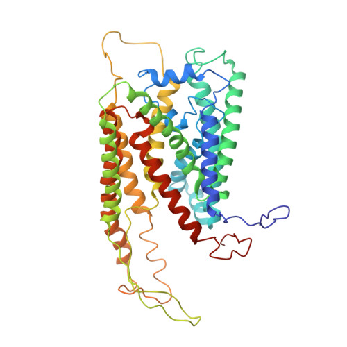
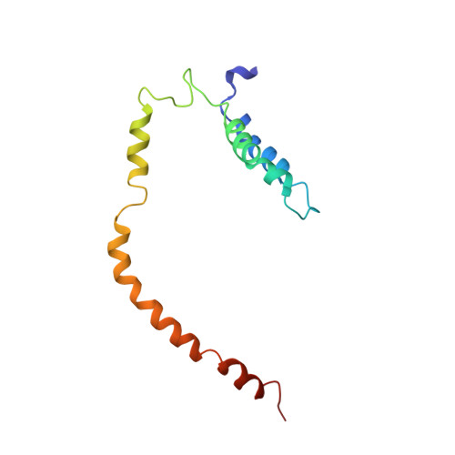
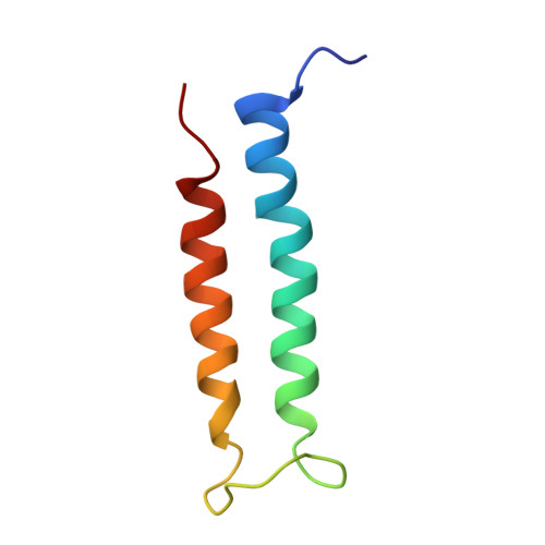
    The biogenesis of polytopic membrane proteins occurs co-translationally on ribosomes that are tightly bound to a membrane-embedded protein-conducting channel: the Sec-complex. The path that is followed by nascent proteins inside the ribosome and the Sec-complex is relatively well established; however, it is not clear what the fate of the N-terminal transmembrane domains (TMDs) of polytopic membrane proteins is when the C-terminal TMDs domains are not yet synthesized. Here, we present the sub-nanometer cryo-electron microscopy structure of an in vivo generated ribosome-SecY complex that carries a membrane insertion intermediate of proteorhodopsin (PR). The structure reveals a pre-opened Sec-complex and the first two TMDs of PR already outside the SecY complex directly in front of its proposed lateral gate. Thus, our structure is in agreement with positioning of N-terminal TMDs at the periphery of SecY, and in addition, it provides clues for the molecular mechanism underlying membrane protein topogenesis.


  • Organizational Affiliation

    Department of Biochemistry, Gene Center and Center for integrated Protein Science Munich, Feodor-Lynen-Strasse 25, University of Munich, 81377 Munich, Germany.


Macromolecules
Find similar proteins by:  (by identity cutoff)  |  3D Structure
Entity ID: 1
MoleculeChains Sequence LengthOrganismDetailsImage
PROTEIN TRANSLOCASE SUBUNIT SECY443Escherichia coliMutation(s): 0 
Membrane Entity: Yes 
UniProt
Find proteins for P0AGA2 (Escherichia coli (strain K12))
Explore P0AGA2 
Go to UniProtKB:  P0AGA2
Entity Groups  
Sequence Clusters30% Identity50% Identity70% Identity90% Identity95% Identity100% Identity
UniProt GroupP0AGA2
Sequence Annotations
Expand
  • Reference Sequence
Find similar proteins by:  (by identity cutoff)  |  3D Structure
Entity ID: 2
MoleculeChains Sequence LengthOrganismDetailsImage
PROTEIN TRANSLOCASE SUBUNIT SECE116Escherichia coliMutation(s): 0 
Membrane Entity: Yes 
UniProt
Find proteins for B7MIW7 (Escherichia coli O45:K1 (strain S88 / ExPEC))
Explore B7MIW7 
Go to UniProtKB:  B7MIW7
Entity Groups  
Sequence Clusters30% Identity50% Identity70% Identity90% Identity95% Identity100% Identity
UniProt GroupB7MIW7
Sequence Annotations
Expand
  • Reference Sequence
Find similar proteins by:  (by identity cutoff)  |  3D Structure
Entity ID: 3
MoleculeChains Sequence LengthOrganismDetailsImage
GREEN-LIGHT ABSORBING PROTEORHODOPSINC [auth Z]69Escherichia coliMutation(s): 0 
Membrane Entity: Yes 
UniProt
Find proteins for Q9F7P4 (Gamma-proteobacterium EBAC31A08)
Explore Q9F7P4 
Go to UniProtKB:  Q9F7P4
Entity Groups  
Sequence Clusters30% Identity50% Identity70% Identity90% Identity95% Identity100% Identity
UniProt GroupQ9F7P4
Sequence Annotations
Expand
  • Reference Sequence
Experimental Data & Validation

Experimental Data

  • Method: ELECTRON MICROSCOPY
  • Resolution: 8.00 Å
  • Aggregation State: PARTICLE 
  • Reconstruction Method: SINGLE PARTICLE 
EM Software:
TaskSoftware PackageVersion
RECONSTRUCTIONSPIDER

Structure Validation

View Full Validation Report


Entry History 

Deposition Data

Revision History  (Full details and data files)

  • Version 1.0: 2015-08-19
    Type: Initial release
  • Version 1.1: 2017-08-30
    Changes: Data collection