4E1K

GlmU in complex with a Quinazoline Compound


Experimental Data Snapshot

  • Method: X-RAY DIFFRACTION
  • Resolution: 2.00 Å
  • R-Value Free: 0.284 
  • R-Value Work: 0.245 
  • R-Value Observed: 0.247 

wwPDB Validation   3D Report Full Report


Ligand Structure Quality Assessment 


This is version 1.1 of the entry. See complete history


Literature

An aminoquinazoline inhibitor of the essential bacterial cell wall synthetic enzyme GlmU has a unique non-protein-kinase-like binding mode.

Larsen, N.A.Nash, T.J.Morningstar, M.Shapiro, A.B.Joubran, C.Blackett, C.J.Patten, A.D.Boriack-Sjodin, P.A.Doig, P.

(2012) Biochem J 446: 405-413

  • DOI: https://doi.org/10.1042/BJ20120596
  • Primary Citation of Related Structures:  
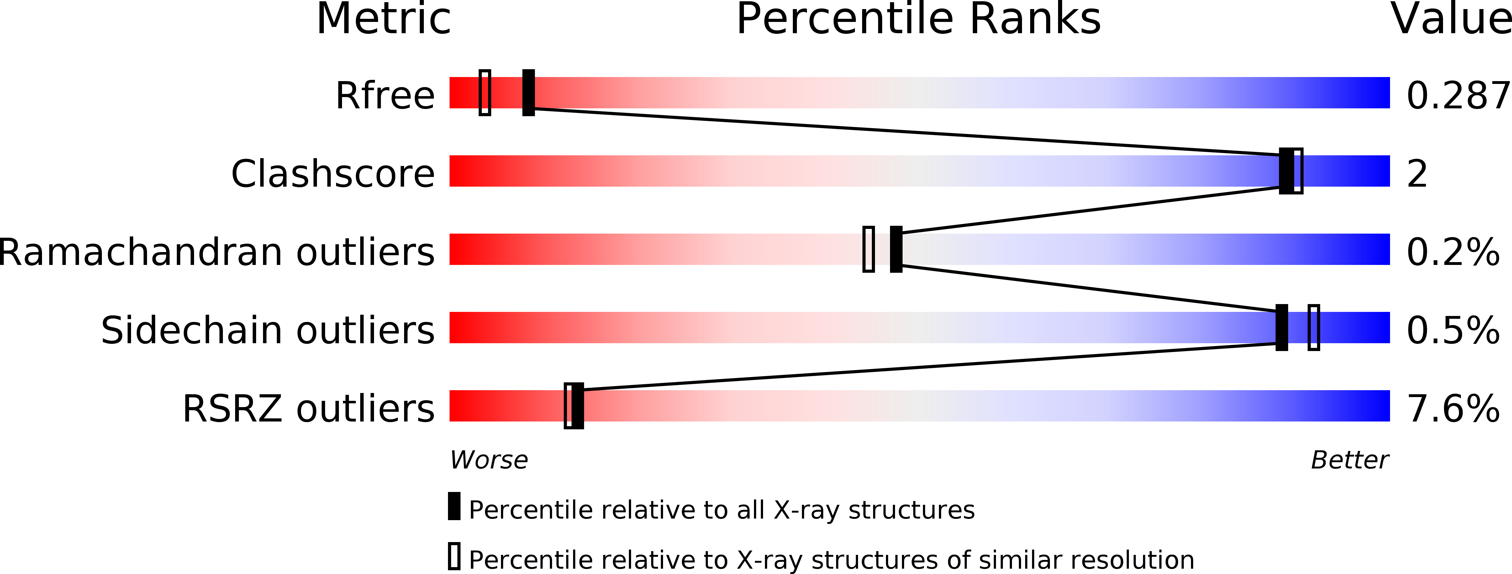
    4E1K

  • PubMed Abstract: 

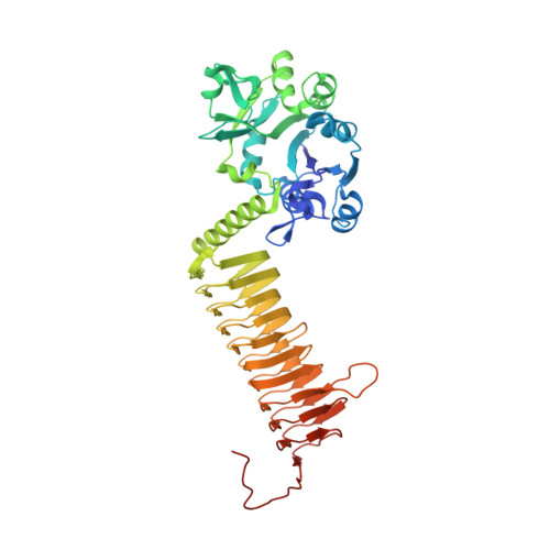
    GlmU is a bifunctional enzyme with acetyltransferase and uridyltransferase activities, and is essential for the biosynthesis of the bacterial cell wall. Inhibition results in a loss of cell viability. GlmU is therefore considered a potential target for novel antibacterial agents. A HTS (high-throughput screen) identified a series of aminoquinazolines with submicromolar potency against the uridyltransferase reaction. Biochemical and biophysical characterization showed competition with UTP binding. We determined the crystal structure of a representative aminoquinazoline bound to the Haemophilus influenzae isoenzyme at a resolution of 2.0 Å. The inhibitor occupies part of the UTP site, skirts the outer perimeter of the GlcNAc1-P (N-acetylglucosamine-1-phosphate) pocket and anchors a hydrophobic moiety into a lipophilic pocket. Our SAR (structure-activity relationship) analysis shows that all of these interactions are essential for inhibitory activity in this series. The crystal structure suggests that the compound would block binding of UTP and lock GlmU in an apo-enzyme-like conformation, thus interfering with its enzymatic activity. Our lead generation effort provides ample scope for further optimization of these compounds for antibacterial drug discovery.


  • Organizational Affiliation

    Discovery Sciences Unit, AstraZeneca R&D Boston, Waltham, MA 02451, USA.


Macromolecules
Find similar proteins by:  (by identity cutoff)  |  3D Structure
Entity ID: 1
MoleculeChains Sequence LengthOrganismDetailsImage
Bifunctional protein GlmU456Haemophilus influenzae Rd KW20Mutation(s): 0 
Gene Names: glmUHI_0642
EC: 2.7.7.23 (PDB Primary Data), 2.3.1.157 (PDB Primary Data)
UniProt
Find proteins for P43889 (Haemophilus influenzae (strain ATCC 51907 / DSM 11121 / KW20 / Rd))
Explore P43889 
Go to UniProtKB:  P43889
Entity Groups  
Sequence Clusters30% Identity50% Identity70% Identity90% Identity95% Identity100% Identity
UniProt GroupP43889
Sequence Annotations
Expand
  • Reference Sequence
Small Molecules
Binding Affinity Annotations 
IDSourceBinding Affinity
0N5 Binding MOAD:  4E1K Kd: 500 (nM) from 1 assay(s)
PDBBind:  4E1K Kd: 500 (nM) from 1 assay(s)
Experimental Data & Validation

Experimental Data

  • Method: X-RAY DIFFRACTION
  • Resolution: 2.00 Å
  • R-Value Free: 0.284 
  • R-Value Work: 0.245 
  • R-Value Observed: 0.247 
  • Space Group: H 3 2
Unit Cell:
Length ( Å )Angle ( ˚ )
a = 109.028α = 90
b = 109.028β = 90
c = 323.602γ = 120
Software Package:
Software NamePurpose
CrystalCleardata collection
AMoREphasing
REFMACrefinement
CrystalCleardata reduction
CrystalCleardata scaling

Structure Validation

View Full Validation Report


Ligand Structure Quality Assessment 


Entry History 

Deposition Data

Revision History  (Full details and data files)

  • Version 1.0: 2013-05-29
    Type: Initial release
  • Version 1.1: 2024-02-28
    Changes: Data collection, Database references, Derived calculations