3OVJ

Structure of an amyloid forming peptide KLVFFA from amyloid beta in complex with orange G


Experimental Data Snapshot

  • Method: X-RAY DIFFRACTION
  • Resolution: 1.80 Å
  • R-Value Free: 0.220 
  • R-Value Work: 0.205 
  • R-Value Observed: 0.207 

wwPDB Validation   3D Report Full Report


Ligand Structure Quality Assessment 


This is version 1.2 of the entry. See complete history


Literature

Towards a pharmacophore for amyloid.

Landau, M.Sawaya, M.R.Faull, K.F.Laganowsky, A.Jiang, L.Sievers, S.A.Liu, J.Barrio, J.R.Eisenberg, D.

(2011) PLoS Biol 9: e1001080-e1001080

  • DOI: https://doi.org/10.1371/journal.pbio.1001080
  • Primary Citation of Related Structures:  
    3OVJ, 3OVL

  • PubMed Abstract: 

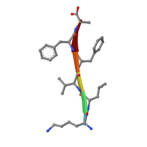
    Diagnosing and treating Alzheimer's and other diseases associated with amyloid fibers remains a great challenge despite intensive research. To aid in this effort, we present atomic structures of fiber-forming segments of proteins involved in Alzheimer's disease in complex with small molecule binders, determined by X-ray microcrystallography. The fiber-like complexes consist of pairs of β-sheets, with small molecules binding between the sheets, roughly parallel to the fiber axis. The structures suggest that apolar molecules drift along the fiber, consistent with the observation of nonspecific binding to a variety of amyloid proteins. In contrast, negatively charged orange-G binds specifically to lysine side chains of adjacent sheets. These structures provide molecular frameworks for the design of diagnostics and drugs for protein aggregation diseases.


  • Organizational Affiliation

    Howard Hughes Medical Institute, UCLA-DOE Institute for Genomics and Proteomics, Department of Biological Chemistry, University of California, Los Angeles, California, United States of America.


Macromolecules

Find similar proteins by:  Sequence   |   3D Structure  

Entity ID: 1
MoleculeChains Sequence LengthOrganismDetailsImage
KLVFFA hexapeptide segment from Amyloid beta
A, B, C, D
6synthetic constructMutation(s): 0 
UniProt & NIH Common Fund Data Resources
Find proteins for P05067 (Homo sapiens)
Explore P05067 
Go to UniProtKB:  P05067
PHAROS:  P05067
GTEx:  ENSG00000142192 
Entity Groups  
UniProt GroupP05067
Sequence Annotations
Expand
  • Reference Sequence
Small Molecules
Ligands 1 Unique
IDChains Name / Formula / InChI Key2D Diagram3D Interactions
ORA
Query on ORA

Download Ideal Coordinates CCD File 
E [auth B],
F [auth D]
7-hydroxy-8-[(E)-phenyldiazenyl]naphthalene-1,3-disulfonic acid
C16 H12 N2 O7 S2
MPVDXIMFBOLMNW-ISLYRVAYSA-N
Experimental Data & Validation

Experimental Data

  • Method: X-RAY DIFFRACTION
  • Resolution: 1.80 Å
  • R-Value Free: 0.220 
  • R-Value Work: 0.205 
  • R-Value Observed: 0.207 
  • Space Group: P 1
Unit Cell:
Length ( Å )Angle ( ˚ )
a = 9.536α = 62.28
b = 26.008β = 88.59
c = 25.803γ = 88.45
Software Package:
Software NamePurpose
SCALEPACKdata scaling
PHASERphasing
REFMACrefinement
PDB_EXTRACTdata extraction
ADSCdata collection
DENZOdata reduction

Structure Validation

View Full Validation Report


Ligand Structure Quality Assessment 


Entry History 

Deposition Data

Revision History  (Full details and data files)

  • Version 1.0: 2011-07-06
    Type: Initial release
  • Version 1.1: 2011-07-13
    Changes: Version format compliance
  • Version 1.2: 2024-02-21
    Changes: Data collection, Database references, Derived calculations