3K7D

C-terminal (adenylylation) domain of E.coli Glutamine Synthetase Adenylyltransferase


Experimental Data Snapshot

  • Method: X-RAY DIFFRACTION
  • Resolution: 2.40 Å
  • R-Value Free: 0.235 
  • R-Value Work: 0.183 
  • R-Value Observed: 0.185 

wwPDB Validation   3D Report Full Report


This is version 1.3 of the entry. See complete history


Literature

Structure of the Adenylylation Domain of E. coli Glutamine Synthetase Adenylyl Transferase: Evidence for Gene Duplication and Evolution of a New Active Site.

Xu, Y.Carr, P.D.Vasudevan, S.G.Ollis, D.L.

(2010) J Mol Biol 396: 773-784

  • DOI: https://doi.org/10.1016/j.jmb.2009.12.011
  • Primary Citation of Related Structures:  
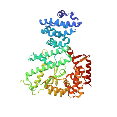
    3K7D

  • PubMed Abstract: 

    The X-ray structure of the C-terminal fragment, containing residues 449-946, of Escherichia coli glutamine synthetase adenylyl transferase (ATase) has been determined. ATase is part of the cascade that regulates the enzymatic activity of E. coli glutamine synthetase, a key component of the cell's machinery for the uptake of ammonia. It has two enzymatic activities, adenylyl removase (AR) and adenylyl transferase (AT), which are located in distinct catalytic domains that are separated by a regulatory (R) domain. We previously reported the three-dimensional structure of the AR domain (residues 1-440). The present structure contains both the R and AT domains. AR and AT share 24% sequence identity and also contain the beta-polymerase motif that is characteristic of many nucleotidylyl transferase enzymes. The structures overlap with an rmsd of 2.4 A when the superhelical R domain is omitted. A model for the complete ATase molecule is proposed, along with some refinements of domain boundaries. A rather more speculative model for the complex of ATase with glutamine synthetase and the nitrogen signal transduction protein PII is also presented.


  • Organizational Affiliation

    Structural Biology Division, The Walter and Eliza Hall Institute of Medical Research, Parkville, Australia.


Macromolecules
Find similar proteins by:  (by identity cutoff)  |  3D Structure
Entity ID: 1
MoleculeChains Sequence LengthOrganismDetailsImage
Glutamate-ammonia-ligase adenylyltransferase
A, B
498Escherichia coliMutation(s): 0 
Gene Names: b3053glnEJW3025
EC: 2.7.7.42
UniProt
Find proteins for P30870 (Escherichia coli (strain K12))
Explore P30870 
Go to UniProtKB:  P30870
Entity Groups  
Sequence Clusters30% Identity50% Identity70% Identity90% Identity95% Identity100% Identity
UniProt GroupP30870
Sequence Annotations
Expand
  • Reference Sequence
Small Molecules
Ligands 1 Unique
IDChains Name / Formula / InChI Key2D Diagram3D Interactions
SO4
Query on SO4

Download Ideal Coordinates CCD File 
C [auth A]
D [auth A]
E [auth A]
F [auth A]
G [auth A]
C [auth A],
D [auth A],
E [auth A],
F [auth A],
G [auth A],
H [auth A],
I [auth A],
J [auth A],
K [auth B],
L [auth B],
M [auth B],
N [auth B],
O [auth B]
SULFATE ION
O4 S
QAOWNCQODCNURD-UHFFFAOYSA-L
Experimental Data & Validation

Experimental Data

  • Method: X-RAY DIFFRACTION
  • Resolution: 2.40 Å
  • R-Value Free: 0.235 
  • R-Value Work: 0.183 
  • R-Value Observed: 0.185 
  • Space Group: P 21 21 21
Unit Cell:
Length ( Å )Angle ( ˚ )
a = 72.542α = 90
b = 122.862β = 90
c = 143.618γ = 90
Software Package:
Software NamePurpose
DENZOdata reduction
SCALEPACKdata scaling
PHASERphasing
REFMACrefinement
PDB_EXTRACTdata extraction
ADSCdata collection

Structure Validation

View Full Validation Report


Entry History 

Deposition Data

Revision History  (Full details and data files)

  • Version 1.0: 2009-12-15
    Type: Initial release
  • Version 1.1: 2011-07-13
    Changes: Advisory, Version format compliance
  • Version 1.2: 2017-11-01
    Changes: Refinement description
  • Version 1.3: 2023-09-06
    Changes: Data collection, Database references, Derived calculations, Refinement description