3HA8

THE COMPLEX STRUCTURE OF THE MAP KINASE P38/Compound 14b


Experimental Data Snapshot

  • Method: X-RAY DIFFRACTION
  • Resolution: 2.48 Å
  • R-Value Free: 0.248 
  • R-Value Work: 0.215 
  • R-Value Observed: 0.217 

wwPDB Validation   3D Report Full Report


This is version 1.2 of the entry. See complete history


Literature

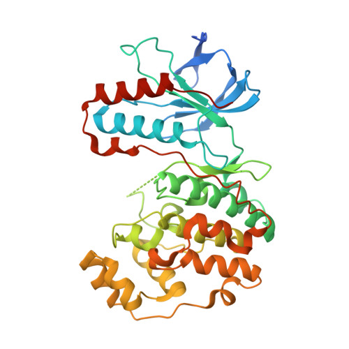
Design, synthesis and selection of DNA-encoded small-molecule libraries.

Clark, M.A.Acharya, R.A.Arico-Muendel, C.C.Belyanskaya, S.L.Benjamin, D.R.Carlson, N.R.Centrella, P.A.Chiu, C.H.Creaser, S.P.Cuozzo, J.W.Davie, C.P.Ding, Y.Franklin, G.J.Franzen, K.D.Gefter, M.L.Hale, S.P.Hansen, N.J.Israel, D.I.Jiang, J.Kavarana, M.J.Kelley, M.S.Kollmann, C.S.Li, F.Lind, K.Mataruse, S.Medeiros, P.F.Messer, J.A.Myers, P.O'Keefe, H.Oliff, M.C.Rise, C.E.Satz, A.L.Skinner, S.R.Svendsen, J.L.Tang, L.van Vloten, K.Wagner, R.W.Yao, G.Zhao, B.Morgan, B.A.

(2009) Nat Chem Biol 5: 647-654

  • DOI: https://doi.org/10.1038/nchembio.211
  • Primary Citation of Related Structures:  
    3HA6, 3HA8

  • PubMed Abstract: 

    Biochemical combinatorial techniques such as phage display, RNA display and oligonucleotide aptamers have proven to be reliable methods for generation of ligands to protein targets. Adapting these techniques to small synthetic molecules has been a long-sought goal. We report the synthesis and interrogation of an 800-million-member DNA-encoded library in which small molecules are covalently attached to an encoding oligonucleotide. The library was assembled by a combination of chemical and enzymatic synthesis, and interrogated by affinity selection. We describe methods for the selection and deconvolution of the chemical display library, and the discovery of inhibitors for two enzymes: Aurora A kinase and p38 MAP kinase.


  • Organizational Affiliation

    Praecis Pharmaceuticals, Waltham, Massachusetts, USA


Macromolecules
Find similar proteins by:  (by identity cutoff)  |  3D Structure
Entity ID: 1
MoleculeChains Sequence LengthOrganismDetailsImage
Mitogen-activated protein kinase 14379Homo sapiensMutation(s): 0 
Gene Names: CSBPCSBP1CSBP2CSPB1MAPK14MXI2P38
EC: 2.7.11.24
UniProt & NIH Common Fund Data Resources
Find proteins for Q16539 (Homo sapiens)
Explore Q16539 
Go to UniProtKB:  Q16539
PHAROS:  Q16539
GTEx:  ENSG00000112062 
Entity Groups  
Sequence Clusters30% Identity50% Identity70% Identity90% Identity95% Identity100% Identity
UniProt GroupQ16539
Sequence Annotations
Expand
  • Reference Sequence
Small Molecules
Ligands 1 Unique
IDChains Name / Formula / InChI Key2D Diagram3D Interactions
5JZ
Query on 5JZ

Download Ideal Coordinates CCD File 
B [auth A]N~2~-{4-[6-(3,4-dihydroquinolin-1(2H)-ylcarbonyl)-1H-benzimidazol-1-yl]-6-ethoxy-1,3,5-triazin-2-yl}-3-(2,2-dimethyl-4H-1,3-benzodioxin-6-yl)-N-methyl-L-alaninamide
C36 H38 N8 O5
IIOQBYZRVQSNSJ-MHZLTWQESA-N
Binding Affinity Annotations 
IDSourceBinding Affinity
5JZ BindingDB:  3HA8 EC50: 8 (nM) from 1 assay(s)
Experimental Data & Validation

Experimental Data

  • Method: X-RAY DIFFRACTION
  • Resolution: 2.48 Å
  • R-Value Free: 0.248 
  • R-Value Work: 0.215 
  • R-Value Observed: 0.217 
  • Space Group: P 21 21 21
Unit Cell:
Length ( Å )Angle ( ˚ )
a = 46.222α = 90
b = 87.934β = 90
c = 121.501γ = 90
Software Package:
Software NamePurpose
REFMACrefinement

Structure Validation

View Full Validation Report


Entry History 

Deposition Data

Revision History  (Full details and data files)

  • Version 1.0: 2009-08-04
    Type: Initial release
  • Version 1.1: 2011-07-13
    Changes: Advisory, Refinement description, Version format compliance
  • Version 1.2: 2023-09-06
    Changes: Data collection, Database references, Derived calculations, Refinement description