3RMN

Human Thrombin in complex with MI341


Experimental Data Snapshot

  • Method: X-RAY DIFFRACTION
  • Resolution: 1.78 Å
  • R-Value Free: 0.185 
  • R-Value Work: 0.154 
  • R-Value Observed: 0.156 

wwPDB Validation   3D Report Full Report


This is version 1.5 of the entry. See complete history


Literature

Ligand binding stepwise disrupts water network in thrombin: enthalpic and entropic changes reveal classical hydrophobic effect

Biela, A.Sielaff, F.Terwesten, F.Heine, A.Steinmetzer, T.Klebe, G.

(2012) J Med Chem 55: 6094-6110

  • DOI: https://doi.org/10.1021/jm300337q
  • Primary Citation of Related Structures:  
    3RLW, 3RLY, 3RM0, 3RM2, 3RML, 3RMM, 3RMN, 3RMO, 3T5F, 3UWJ

  • PubMed Abstract: 

    Well-ordered water molecules are displaced from thrombin's hydrophobic S3/4-pocket by P3-varied ligands (Gly, d-Ala, d-Val, d-Leu to d-Cha with increased hydrophobicity and steric requirement). Two series with 2-(aminomethyl)-5-chlorobenzylamide and 4-amidinobenzylamide at P1 were examined by ITC and crystallography. Although experiencing different interactions in S1, they display almost equal potency. For both scaffolds the terminal benzylsulfonyl substituent differs in binding, whereas the increasingly bulky P3-groups address S3/4 pocket similarly. Small substituents leave the solvation pattern unperturbed as found in the uncomplexed enzyme while increasingly larger ones stepwise displace the waters. Medium-sized groups show patterns with partially occupied waters. The overall 40-fold affinity enhancement correlates with water displacement and growing number of van der Waals contacts and is mainly attributed to favorable entropy. Both Gly derivatives deviate from the series and adopt different binding modes. Nonetheless, their thermodynamic signatures are virtually identical with the homologous d-Ala derivatives. Accordingly, unchanged thermodynamic profiles are no reliable indicator for conserved binding modes.


  • Organizational Affiliation

    Department of Pharmaceutical Chemistry, Philipps University Marburg, Marbacher Weg 6, 35032 Marburg, Germany.


Macromolecules
Find similar proteins by:  (by identity cutoff)  |  3D Structure
Entity ID: 1
MoleculeChains Sequence LengthOrganismDetailsImage
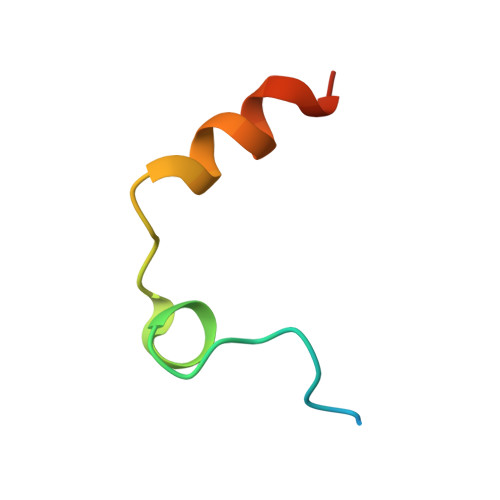
Thrombin Light ChainA [auth L]36Homo sapiensMutation(s): 0 
EC: 3.4.21.5
UniProt & NIH Common Fund Data Resources
Find proteins for P00734 (Homo sapiens)
Explore P00734 
Go to UniProtKB:  P00734
PHAROS:  P00734
GTEx:  ENSG00000180210 
Entity Groups  
Sequence Clusters30% Identity50% Identity70% Identity90% Identity95% Identity100% Identity
UniProt GroupP00734
Sequence Annotations
Expand
  • Reference Sequence
Find similar proteins by:  (by identity cutoff)  |  3D Structure
Entity ID: 2
MoleculeChains Sequence LengthOrganismDetailsImage
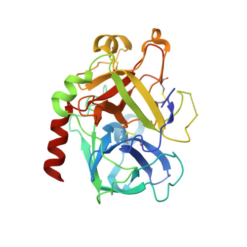
Thrombin Heavy ChainB [auth H]259Homo sapiensMutation(s): 0 
EC: 3.4.21.5
UniProt & NIH Common Fund Data Resources
Find proteins for P00734 (Homo sapiens)
Explore P00734 
Go to UniProtKB:  P00734
PHAROS:  P00734
GTEx:  ENSG00000180210 
Entity Groups  
Sequence Clusters30% Identity50% Identity70% Identity90% Identity95% Identity100% Identity
UniProt GroupP00734
Sequence Annotations
Expand
  • Reference Sequence

Find similar proteins by:  Sequence   |   3D Structure  

Entity ID: 3
MoleculeChains Sequence LengthOrganismDetailsImage
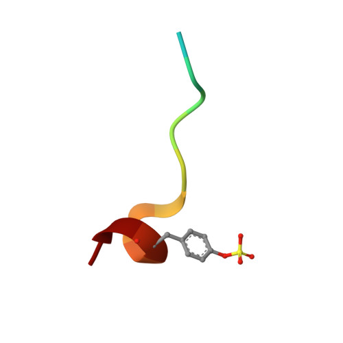
Hirudin variant-2C [auth I]13Hirudo medicinalisMutation(s): 0 
UniProt
Find proteins for P09945 (Hirudo medicinalis)
Explore P09945 
Go to UniProtKB:  P09945
Entity Groups  
Sequence Clusters30% Identity50% Identity70% Identity90% Identity95% Identity100% Identity
UniProt GroupP09945
Sequence Annotations
Expand
  • Reference Sequence
Small Molecules
Ligands 5 Unique
IDChains Name / Formula / InChI Key2D Diagram3D Interactions
M41
Query on M41

Download Ideal Coordinates CCD File 
E [auth H]N-(benzylsulfonyl)-D-valyl-N-[2-(aminomethyl)-5-chlorobenzyl]-L-prolinamide
C25 H33 Cl N4 O4 S
PWCVAVZHMMVMMM-XZOQPEGZSA-N
NAG
Query on NAG

Download Ideal Coordinates CCD File 
D [auth H]2-acetamido-2-deoxy-beta-D-glucopyranose
C8 H15 N O6
OVRNDRQMDRJTHS-FMDGEEDCSA-N
PO4
Query on PO4

Download Ideal Coordinates CCD File 
F [auth H]PHOSPHATE ION
O4 P
NBIIXXVUZAFLBC-UHFFFAOYSA-K
GOL
Query on GOL

Download Ideal Coordinates CCD File 
G [auth H],
H
GLYCEROL
C3 H8 O3
PEDCQBHIVMGVHV-UHFFFAOYSA-N
NA
Query on NA

Download Ideal Coordinates CCD File 
I [auth H],
J [auth H]
SODIUM ION
Na
FKNQFGJONOIPTF-UHFFFAOYSA-N
Modified Residues  1 Unique
IDChains TypeFormula2D DiagramParent
TYS
Query on TYS
C [auth I]L-PEPTIDE LINKINGC9 H11 N O6 STYR
Binding Affinity Annotations 
IDSourceBinding Affinity
M41 BindingDB:  3RMN Ki: 0.8 (nM) from 1 assay(s)
Biologically Interesting Molecules (External Reference) 1 Unique
Experimental Data & Validation

Experimental Data

  • Method: X-RAY DIFFRACTION
  • Resolution: 1.78 Å
  • R-Value Free: 0.185 
  • R-Value Work: 0.154 
  • R-Value Observed: 0.156 
  • Space Group: C 1 2 1
Unit Cell:
Length ( Å )Angle ( ˚ )
a = 69.9α = 90
b = 71.1β = 100.6
c = 72.9γ = 90
Software Package:
Software NamePurpose
MAR345dtbdata collection
PHASERphasing
PHENIXrefinement
HKL-2000data reduction
HKL-2000data scaling

Structure Validation

View Full Validation Report


Entry History 

Deposition Data

Revision History  (Full details and data files)

  • Version 1.0: 2012-04-25
    Type: Initial release
  • Version 1.1: 2012-12-12
    Changes: Other
  • Version 1.2: 2016-07-13
    Changes: Database references
  • Version 1.3: 2020-07-29
    Type: Remediation
    Reason: Carbohydrate remediation
    Changes: Data collection, Derived calculations, Structure summary
  • Version 1.4: 2023-11-01
    Changes: Data collection, Database references, Refinement description, Structure summary
  • Version 1.5: 2023-12-06
    Changes: Data collection