Hydrogen exchange study on the hydroxyl groups of serine and threonine residues in proteins and structure refinement using NOE restraints with polar side-chain groups
Takeda, M., Jee, J., Ono, A.M., Terauchi, T., Kainosho, M.(2011) J Am Chem Soc 133: 17420-17427
- PubMed: 21955241
- DOI: https://doi.org/10.1021/ja206799v
- Primary Citation of Related Structures:
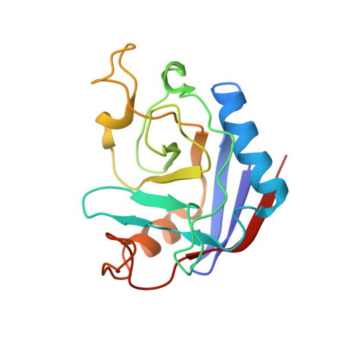
2RS4 - PubMed Abstract:
We recently developed new NMR methods for monitoring the hydrogen exchange rates of tyrosine hydroxyl (Tyr-OH) and cysteine sulfhydryl (Cys-SH) groups in proteins. These methods facilitate the identification of slowly exchanging polar side-chain protons in proteins, which serve as sources of NOE restraints for protein structure refinement. Here, we have extended the methods for monitoring the hydrogen exchange rates of the OH groups of serine (Ser) and threonine (Thr) residues in an 18.2 kDa protein, EPPIb, and thus demonstrated the usefulness of NOE restraints with slowly exchanging OH protons for refining the protein structure. The slowly exchanging Ser/Thr-OH groups were readily identified by monitoring the (13)C(β)-NMR signals in an H(2)O/D(2)O (1:1) mixture, for the protein containing Ser/Thr residues with (13)C, (2)H-double labels at their β carbons. Under these circumstances, the OH groups exist in equilibrium between the protonated and deuterated isotopomers, and the (13)C(β) peaks of the two species are resolved when their exchange rate is slower than the time scale of the isotope shift effect. In the case of EPPIb dissolved in 50 mM sodium phosphate buffer (pH 7.5) at 40 °C, one Ser and four Thr residues were found to have slowly exchanging hydroxyl groups (k(ex) < ~40 s(-1)). With the information for the slowly exchanging Ser/Thr-OH groups in hand, we could collect additional NOE restraints for EPPIb, thereby making a unique and important contribution toward defining the spatial positions of the OH protons, and thus the hydrogen-bonding acceptor atoms.
Organizational Affiliation:
Structural Biology Research Center, Graduate School of Science, Nagoya University, Nagoya 464-8602, Japan.