2BYV

Structure of the cAMP responsive exchange factor Epac2 in its auto- inhibited state


Experimental Data Snapshot

  • Method: X-RAY DIFFRACTION
  • Resolution: 2.70 Å
  • R-Value Free: 0.297 
  • R-Value Work: 0.245 
  • R-Value Observed: 0.248 

wwPDB Validation   3D Report Full Report


This is version 1.3 of the entry. See complete history


Literature

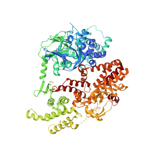
Structure of the Cyclic-AMP Responsive Exchange Factor Epac2 in its Auto-Inhibited State

Rehmann, H.Das, J.Knipscheer, P.Wittinghofer, A.Bos, J.L.

(2006) Nature 439: 625

  • DOI: https://doi.org/10.1038/nature04468
  • Primary Citation of Related Structures:  
    2BYV

  • PubMed Abstract: 

    Epac proteins (exchange proteins directly activated by cAMP) are guanine-nucleotide-exchange factors (GEFs) for the small GTP-binding proteins Rap1 and Rap2 that are directly regulated by the second messenger cyclic AMP and function in the control of diverse cellular processes, including cell adhesion and insulin secretion. Here we report the three-dimensional structure of full-length Epac2, a 110-kDa protein that contains an amino-terminal regulatory region with two cyclic-nucleotide-binding domains and a carboxy-terminal catalytic region. The structure was solved in the absence of cAMP and shows the auto-inhibited state of Epac. The regulatory region is positioned with respect to the catalytic region by a rigid, tripartite beta-sheet-like structure we refer to as the 'switchboard' and an ionic interaction we call the 'ionic latch'. As a consequence of this arrangement, the access of Rap to the catalytic site is sterically blocked. Mutational analysis suggests a model for cAMP-induced Epac activation with rigid body movement of the regulatory region, the features of which are universally conserved in cAMP-regulated proteins.


  • Organizational Affiliation

    Department of Physiological Chemistry and Centre for Biomedical Genetics, University Medical Center Utrecht, Universiteitsweg 100, 3584 CG Utrecht, The Netherlands.


Macromolecules
Find similar proteins by:  (by identity cutoff)  |  3D Structure
Entity ID: 1
MoleculeChains Sequence LengthOrganismDetailsImage
RAP GUANINE NUCLEOTIDE EXCHANGE FACTOR 4A [auth E]999Mus musculusMutation(s): 0 
UniProt & NIH Common Fund Data Resources
Find proteins for Q9EQZ6 (Mus musculus)
Explore Q9EQZ6 
Go to UniProtKB:  Q9EQZ6
IMPC:  MGI:1917723
Entity Groups  
Sequence Clusters30% Identity50% Identity70% Identity90% Identity95% Identity100% Identity
UniProt GroupQ9EQZ6
Sequence Annotations
Expand
  • Reference Sequence
Experimental Data & Validation

Experimental Data

  • Method: X-RAY DIFFRACTION
  • Resolution: 2.70 Å
  • R-Value Free: 0.297 
  • R-Value Work: 0.245 
  • R-Value Observed: 0.248 
  • Space Group: P 21 21 21
Unit Cell:
Length ( Å )Angle ( ˚ )
a = 76.406α = 90
b = 97.038β = 90
c = 172.101γ = 90
Software Package:
Software NamePurpose
REFMACrefinement
XDSdata reduction
XSCALEdata scaling
MOLREPphasing

Structure Validation

View Full Validation Report


Entry History 

Deposition Data

Revision History  (Full details and data files)

  • Version 1.0: 2006-02-02
    Type: Initial release
  • Version 1.1: 2011-05-08
    Changes: Version format compliance
  • Version 1.2: 2011-07-13
    Changes: Version format compliance
  • Version 1.3: 2023-12-13
    Changes: Data collection, Database references, Other, Refinement description